Inflammatory pigmented paravenous retinochoroidal atrophy
- Select a language for the TTS:
- UK English Female
- UK English Male
- US English Female
- US English Male
- Australian Female
- Australian Male
- Language selected: (auto detect) - EN

Play all audios:

ABSTRACT _Purpose_ To describe an active inflammatory cause of pigmented paravenous retinochoroidal atrophy. _Methods_ A 54-year-old female patient presented with complaints of worsening
visual acuity and poor night vision was examined. Fundus examination was performed and color fundus photographs were taken. In addition to fluorescein angiography, visual field examinations
and electroretinographic tests were performed. Macular evaluation was performed with optical coherence tomography. _Results_ Both fundi showed circumscribed patches of retinochoroidal
atrophy and pigmentation along the retinal veins. She had also marked vitreous cells with snow ball opacities and cystoid macular edema in both eyes. Fluorescein angiography confirmed the
presence of a hyperfluorescence due to widespread paravenous retinal pigment epithelial defect while ICG angiography disclosed hypofluorescence in all phases. The electroretinogram showed
reduced responses especially in the left eye. Visual field tests showed scotomas corresponding with areas of atrophy along the retinal veins. _Conclusions_ This is a report of the findings
in pigmented paravenous retinochoroidal atrophy that is a nonspecific degenerative disease and may occur in association with systemic infections or inflammation. Ocular inflammation with
cystoid macular edema is an unusual manifestation of the disease. SIMILAR CONTENT BEING VIEWED BY OTHERS THE IMPLICATIONS OF SUBRETINAL FLUID IN PACHYCHOROID NEOVASCULOPATHY Article Open
access 18 February 2021 SMALL DOME-SHAPED PIGMENT EPITHELIUM DETACHMENT IN POLYPOIDAL CHOROIDAL VASCULOPATHY: AN UNDER-RECOGNIZED SIGN OF POLYPOIDAL LESIONS ON OPTICAL COHERENCE TOMOGRAPHY?
Article 08 April 2021 NON-EXUDATIVE OCT FINDINGS IN NEOVASCULAR AMD Article 25 November 2024 MAIN Pigmented paravenous retinochoroidal atrophy (PPRA) is an uncommon disease characterised by
paravenous zones of retinal degeneration with bone spicule pigmentation.1,2,3,4,5,6,7,8,9 This is an asymptomatic disease usually detected during routine ophthalmic examination. The
condition is generally bilateral and young adults are most commonly affected. The cause of the disease is unknown although an inflammatory10 or hereditary11 aetiology has been suggested. We
report a case with typical fundus appearance of paravenous pigmented retinochoroidal atrophy accompanied by an active inflammation with cystoid macular edema. CASE REPORT A 54-year-old
woman, first seen in December 1999, presented with complaints of worsening visual acuity and poor night vision for 1.5 years. The review of her medical history indicated that she had
diabetes mellitus and systemic hypertension for 2 years. Ophthalmic examination revealed that her best corrected visual acuity was RE: 8/10 and LE: 3/10. External examination was normal and
extraocular motility was full. Slit-lamp examination disclosed marked cells and snowball opacities in the vitreous of both eyes. Intraocular pressure was 15 mmHg in each eye. Fundus
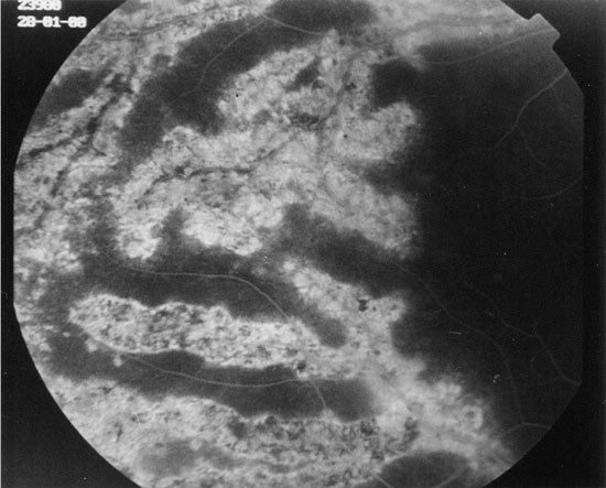
examination showed circumscribed patches of retinochoroidal atrophy and pigmentation along the retinal veins and cystoid macular edema in both eyes. Fluorescein angiography showed diffuse
window defects with hyperfluorescence consistent with retinal pigment epithelial degeneration, blocking of the fluorescence in the areas of pigment clumping along retinal vessels (Figure 1)
and cystoid macular edema in both eyes (Figure 2a, b). Cystoid macular edema was also demonstrated with optical coherence tomography in both eyes (Figure 3). Colour vision with Ishihara
Pseudoisochromatic plates was normal in each eye. Humprey field analyser showed scotomas corresponding with areas of atrophy along the retinal veins. Electrophysiological tests were
performed showing reduced a and b wave amplitudes in rod response (scotopic ERG) and maximal combined response. Pattern VEP showed minimal decrease of amplitude and increase in latency in
the left eye in comparison to the right eye, however the values for both eyes fell within the normal range for a patient of this age. Pattern ERG showed reduced N95 amplitude in the left eye
(3.6 μV in the right and 2.2 μV in the left). PVEP and PERG findings showed ganglion cell involvement in the left eye in comparison to the right eye. There was a subnormal ratio of light
peak/dark trough voltage in electrooculogram suggesting dysfunction in the region of RPE in both eyes. No systemic abnormality was found. Results of laboratory studies including complete
blood cell counts, serum electrolytes, serum protein electrophoresis, erythrocyte sedimentation rate were within normal range. Tuberculin skin test was 15 mm. There was no serologic evidence
of syphilis, toxoplasmosis, systemic lupus eritematosis. Serum antibody levels for HSV I-II, CMV, rubella and measles were all negative. During the follow-up period of 12 months, there was
no change in cystoid macular edema and the fundus appearance although we treated the patient with corticosteroids and acetozolamide. DISCUSSION The diagnosis of PPRA is based on a typical
and characteristic fundus appearance such as: areas of atrophy of the retina, pigment epithelium, and choriocapillaris around the optic disc and along the retinal veins, bone corpuscle
pigment accumulation along the distribution of retinal veins. Fluorescein angiography, electrophysiological tests and visual fields may confirm the diagnosis. The patient described here
showed bilateral cystoid macular edema and perivenous aggregations of pigment clumps associated with peripapillary and radial zones of chorioretinal atrophy. This fundus appearance was
characteristic of pigmented paravenous retinochoroidal atrophy. The association of active inflammation with cystoid macular edema, however was unique in this case. There have been limited
number of reports of active inflammation with this disease. Yamaguchi _et al_ reported a 47-year-old Japanese man who had a progressive degeneration of the retina and choroid along the
retinal veins associated with uveitis of 2 years’ duration.10 Haustrate and Osterhuis reported five patients with PPRA and in one patient they observed signs of active uveitis and
progression of fundus lesions.3 Macular involvement is very rare in this disease. Bilateral macular coloboma was reported by Chen _et al_ in a 23-year-old woman.12 Our case had bilateral
cystoid macular edema which was one of the manifestations of active inflammation, and as far as we are aware this has not been previously reported. Fluorescein angiography gave a better
definition of the lesions as well as cystoid macular edema. Optical coherence tomography revealed large cystoid areas in macular cross-sections. In PPRA it is likely that the fundus lesions
may be slowly progressive4,8,13 and that the fundus resembles retinitis pigmentosa with time. In a few patients, the fundus condition may be progressive and may be detected at an older age.5
In our case during the 9 months follow-up period, we did not observe progression in fundus lesions. Macular edema did not resolve despite treatment with steroids and acetozolamide. The
aetiology of this disease is unknown. However some authors suggest a congenital origin9,11 and others a primary retinal degeneration.1 Chen _et al_ reported a case with bilateral macular
coloboma, PPRA and negative laboratory examinations and he speculated that it was a developmental abnormality in nature.12 Some reports suggest an inflammatory cause. Syphilitic aetiology
has been suggested by Chi Hsin-Hsiang.14 Scheie15 described a case with rubeola retinopathy which progressed to the stage of secondary pigmentary degeneration of the retina and later Foxman
_et al_16 examined the same patient at the age of 36. Our patient did not have a systemic abnormality according to her medical history and laboratory evaluation. The differential diagnosis
includes both chorioretinal degeneration and inflammatory disease that cause chorioretinal atrophy, such as helicoid peripapillary chorioretinal atrophy, proliferating choroiditis, angioid
streaks, gyrate atrophy choroideraemia, Wagner’s dominant vitreoretinal degeneration, sarcoidosis, syphilis, acute retinal necrosis, CMV retinitis, tuberculous disseminated choroiditis,
onchocerciasis, toxoplasmosis and frosted branch angiitis. In conclusion, PPRA is a nonspecific degenerative disease that may occur in association with systemic infections or inflammation.
Ocular inflammation with cystoid macular edema is an unusual manifestation of the disease. REFERENCES * Noble KG, Carr RE . Pigmented paravenous chorioretinal atrophy. _Am J Ophthalmol_
1983; 96: 338–344 Article CAS Google Scholar * Rothberg DS, Cibis GW, Trese M . Paravenous pigmentary retinochoroidal atrophy. _Ann Ophthalmol_ 1984; 16: 643–646 CAS PubMed Google
Scholar * Haustrate FMRJ, Oosterhuis JA . Pigmented paravenous retinochoroidal atrophy. _Doc Ophthalmol_ 1986; 63: 209–237 Article CAS Google Scholar * Klop K, Van Schooneveld MJ .
Pigmented paravenous retinochoroidal atrophy: a nosologic entity?. _Doc Ophthalmol_ 1988; 70: 185–193 Article CAS Google Scholar * Hayasaka S, Shibasaki H, Noda S, Fujii M, Setogawa T .
Pigmented paravenous retinochoroidal atrophy in a 68-year-old man. _Ann Ophthalmol_ 1991; 23: 177–180 CAS PubMed Google Scholar * Parafita M, Diaz A, Torrijos IG, Gomez-Ulla F . Pigmented
paravenous retinochoroidal atrophy. _Optometry and Vision Science_ 1993; 70: 75–78 Article CAS Google Scholar * Pearlman JT, Kamin DF, Kopelow SM, Saxton J . Pigmented paravenous
retinochoroidal atrophy. _Am J Ophthalmol_ 1975; 80: 630–635 Article CAS Google Scholar * Pearlman JT, Heckenlively JR, Bastek JV . Progressive nature of paravenous chorioretinal atrophy.
_Am J Ophthalmol_ 1978; 85: 215–217 Article CAS Google Scholar * Small KW, Anderson WB . Pigmented paravenous retinochoroidal atrophy discordant expression in monozygotic twins. _Arch
Ophthalmol_ 1991; 109: 1408–1410 Article CAS Google Scholar * Yamaguchi K, Hara S, Tanifuji Y, Tamai M . Inflammatory pigmented paravenous retinochoroidal atrophy. _Br J Ophthalmol_ 1989;
73: 463–467 Article CAS Google Scholar * Skalka HW . Hereditary pigmented paravenous retinochoroidal atrophy. _Am J Ophthalmol_ 1979; 87: 286–291 Article CAS Google Scholar * Chen MS,
Yang CH, Huang JS . Bilateral macular coloboma and pigmented paravenous retinochoroidal atrophy. _Br J Ophthalmol_ 1992; 76: 250–251 Article CAS Google Scholar * Limaye SR, Mahmood MA .
Retinal microangiopathy in pigmented paravenous chorioretinal atrophy. _Br J Ophthalmol_ 1987; 71: 757–761 Article CAS Google Scholar * Hsin-Hsiang C . Retinochoroiditis radiata. _Am J
Ophthalmol_ 1948; 31: 1485–1487 Article Google Scholar * Scheie HG, Morse PH . Rubeola retinopathy. _Arch Ophthalmol_ 1972; 88: 341–344 Article CAS Google Scholar * Foxman SG,
Heckenlively JR, Sinclair SH . Rubeola retinopathy and pigmented paravenous retinochoroidal atrophy. _Am J Ophthalmol_ 1985; 99: 605–606 Article CAS Google Scholar Download references
AUTHOR INFORMATION AUTHORS AND AFFILIATIONS * Ankara University School of Medicine Eye Clinic Ankara, Turkey F Batioğlu, L S Atmaca, H Atilla & A Arslanpençe Authors * F Batioğlu View
author publications You can also search for this author inPubMed Google Scholar * L S Atmaca View author publications You can also search for this author inPubMed Google Scholar * H Atilla
View author publications You can also search for this author inPubMed Google Scholar * A Arslanpençe View author publications You can also search for this author inPubMed Google Scholar
CORRESPONDING AUTHOR Correspondence to F Batioğlu. ADDITIONAL INFORMATION This case report was presented at the VIth Mediterranean Ophthalmological Society Congress and VIth Michealson
Symposium May 21–26 2001, Jerusalem, Israel RIGHTS AND PERMISSIONS Reprints and permissions ABOUT THIS ARTICLE CITE THIS ARTICLE Batioğlu, F., Atmaca, L., Atilla, H. _et al._ Inflammatory
pigmented paravenous retinochoroidal atrophy. _Eye_ 16, 81–84 (2002). https://doi.org/10.1038/sj.eye.6700021 Download citation * Published: 05 April 2002 * Issue Date: 01 January 2002 * DOI:
https://doi.org/10.1038/sj.eye.6700021 SHARE THIS ARTICLE Anyone you share the following link with will be able to read this content: Get shareable link Sorry, a shareable link is not
currently available for this article. Copy to clipboard Provided by the Springer Nature SharedIt content-sharing initiative KEYWORDS * Pigmented paravenous retinochoroidal atrophy * ocular
inflammation * cystoid macular edema