Late presentation of severe thyroid eye disease
- Select a language for the TTS:
- UK English Female
- UK English Male
- US English Female
- US English Male
- Australian Female
- Australian Male
- Language selected: (auto detect) - EN
Play all audios:

The presentation of thyroid eye disease is usually closely temporally related to the diagnosis of thyrotoxicosis. A very small percentage of patients with thyroid eye disease develop
sight-threatening disease due to compressive optic neuropathy. We report a case of severe thyroid eye disease with sight loss presenting 14 years after the diagnosis of thyrotoxicosis.
A 57-year-old Caucasian female, who smoked five cigarettes a day, presented with a severe reduction in right visual acuity associated with proptosis. Fourteen years previously, she had been
treated with carbimazole and radioactive iodine for thyrotoxicosis. Subsequent hypothyroidism was treated with thyroxine. She had controlled hypertension and seasonal episodes of bronchitis.
There was a strong family history of thyroid problems with her mother and sister being on replacement thyroxine treatment, although the underlying thyroid condition was unknown. Her
ophthalmological symptoms had started approximately 9 months previously, shortly after the death of her father, initially with right lower lid swelling followed 3 months later by horizontal
diplopia and conjunctival injection. She then developed blurred right vision and she was referred to the local Eye Department where it was felt unlikely that she had thyroid eye disease in
view of the long interval between her thyrotoxicosis and the onset of her eye problems. Three months later, she developed right proptosis with reduced vision and following a CT scan was
started on 100 μg prednisolone reducing to 10 μg over 3 weeks. The left vision deteriorated, and the steroid dosage was increased to a maintenance dose of 25 μg. She was referred to our
service for further opinion, and presented with a corrected Snellen visual acuity of 2/60 in her right and 6/12 in her left eye. She was unable to identify any of the Ishihara test plates
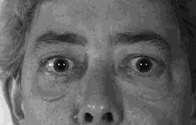
with her right eye and scored only 10 out of 17 with the left eye. There was right proptosis with upper and lower lid retraction and restricted extraocular movements worse on the right side
(Figure 1). The anterior segments were normal. Intraocular pressures increased significantly on upgaze. She had a right afferent pupillary defect, nasal swelling of the right optic disc,
absent spontaneous venous pulsation of both optic discs, and extensive arteriovenous nipping.
Electrodiagnostic tests were reported as consistent with a diagnosis of a right optic neuropathy. The MRI scan showed compression of the right optic nerve at the orbital apex, with bilateral
proptosis more marked on the right side due to asymmetrical enlargement of the extraocular muscles (Figure 2). Blood results showed a neutrophilia, normal haemoglobin and platelet
concentrations with normal blood chemistry. Thyroid function tests had recently shown an elevated free thyroxine concentration with an associated low TSH level and her thyroxine had been
reduced to 75 μg.
MRI scan showing compression of the right optic nerve at the orbital apex.
The patient was started on a 2-day course of intravenous methylprednisolone and a three-wall orbital decompression was advised.
Although the majority of patients with thyroid eye disease usually present very soon after the diagnosis of thyrotoxicosis, it is important to remember that, rarely, late presentation can
occur. Anecdotally, it has been suggested that these patients can often identify a stressful and identifiable precipitating factor. Such an event was noted in our patient. Only 5% of
patients have the complete spectrum of clinical features with eyelid retraction, exophthalmos, extraocular muscle involvement, optic nerve dysfunction and hyperthyroidism.1 The close
temporal relationship of the diagnosis of Graves' disease and thyroid eye disease is well known and it has been found in 81% of patients to develop in the 18 months prior to and after the
diagnosis of thyroid dysfunction.2 It has been reported that 69.5% of patients requiring decompression surgery develop thyroid eye disease in the year before or after diagnosis.3
Ophthalmopathy is rarely diagnosed more than 6 months before the diagnosis of thyrotoxicosis.4 In one report no patient developed eye problems more than 4 years after the diagnosis of
hyperthyroidism.4 It has been suggested that thyroid eye disease develops at a time when thyroid autoimmunity also exists, thus strongly suggesting a common factor in the pathogenesis of
thyroidal and ocular expressions of Graves' disease.5
This patient is a rare case of severe sight-threatening thyroid eye disease presenting 14 years after diagnosis of thyrotoxicosis. While the vast majority of patients who develop thyroid eye
disease will do so in an 18-month period before or after the time of systemic diagnosis, it is important to be aware that sight-threatening thyroid eye disease may present many years after
the initial diagnosis and treatment of thyrotoxicosis. Prompt diagnosis and treatment of this condition is likely to preserve sight.
Anyone you share the following link with will be able to read this content: