Three-dimensional imaging of localized surface plasmon resonances of metal nanoparticles
- Select a language for the TTS:
- UK English Female
- UK English Male
- US English Female
- US English Male
- Australian Female
- Australian Male
- Language selected: (auto detect) - EN

Play all audios:

ABSTRACT The remarkable optical properties of metal nanoparticles are governed by the excitation of localized surface plasmon resonances (LSPRs). The sensitivity of each LSPR mode, whose
spatial distribution and resonant energy depend on the nanoparticle structure, composition and environment, has given rise to many potential photonic, optoelectronic, catalytic,
photovoltaic, and gas- and bio-sensing applications1,2,3. However, the precise interplay between the three-dimensional (3D) nanoparticle structure and the LSPRs is not always fully
understood and a spectrally sensitive 3D imaging technique is needed to visualize the excitation on the nanometre scale. Here we show that 3D images related to LSPRs of an individual silver
nanocube can be reconstructed through the application of electron energy-loss spectrum imaging4, mapping the excitation across a range of orientations, with a novel combination of
non-negative matrix factorization5,6, compressed sensing7,8 and electron tomography9. Our results extend the idea of substrate-mediated hybridization of dipolar and quadrupolar modes
predicted by theory, simulations, and electron and optical spectroscopy10,11,12, and provide experimental evidence of higher-energy mode hybridization. This work represents an advance both
in the understanding of the optical response of noble-metal nanoparticles and in the probing, analysis and visualization of LSPRs. Access through your institution Buy or subscribe This is a
preview of subscription content, access via your institution ACCESS OPTIONS Access through your institution Subscribe to this journal Receive 51 print issues and online access $199.00 per
year only $3.90 per issue Learn more Buy this article * Purchase on SpringerLink * Instant access to full article PDF Buy now Prices may be subject to local taxes which are calculated during
checkout ADDITIONAL ACCESS OPTIONS: * Log in * Learn about institutional subscriptions * Read our FAQs * Contact customer support SIMILAR CONTENT BEING VIEWED BY OTHERS OPTICAL SPECTRA OF
SILVER CLUSTERS AND NANOPARTICLES FROM 4 TO 923 ATOMS FROM THE TDDFT+_U_ METHOD Article Open access 25 October 2024 LARGE FIELD-OF-VIEW PLASMONIC SCATTERING IMAGING AND SENSING OF
NANOPARTICLES WITH ISOTROPIC POINT-SPREAD-FUNCTION Article Open access 02 June 2025 SIMPLE EXPERIMENTAL PROCEDURES TO DISTINGUISH PHOTOTHERMAL FROM HOT-CARRIER PROCESSES IN PLASMONICS
Article Open access 28 June 2020 REFERENCES * Maier, S. A. & Atwater, H. A. Plasmonics: localization and guiding of electromagnetic energy in metal/dielectric structures. _J. Appl.
Phys._ 98, 011101 (2005) Article ADS Google Scholar * Schuller, J. A. et al. Plasmonics for extreme light concentration and manipulation. _Nature Mater._ 9, 193–204 (2010) Article ADS
CAS Google Scholar * Anker, J. N. et al. Biosensing with plasmonic nanosensors. _Nature Mater._ 7, 442–453 (2008) Article ADS CAS Google Scholar * Jeanguillaume, C. & Colliex, C.
Spectrum-image: the next step in EELS digital acquisition and processing. _Ultramicroscopy_ 28, 252–257 (1989) Article Google Scholar * Paatero, P. & Tapper, U. Positive matrix
factorization: a non-negative factor model with optimal utilization of error estimates of data values. _Environmetrics_ 5, 111–126 (1994) Article Google Scholar * Lee, D. D. & Seung,
H. S. Learning the parts of objects by non-negative matrix factorization. _Nature_ 401, 788–791 (1999) Article ADS CAS PubMed Google Scholar * Donoho, D. L. Compressed sensing. _IEEE
Trans. Inf. Theory_ 52, 1289–1306 (2006) Article MathSciNet Google Scholar * Candes, E. J., Romberg, J. & Tao, T. Robust uncertainty principles: exact signal reconstruction from
highly incomplete frequency information. _IEEE Trans. Inf. Theory_ 52, 489–509 (2006) Article MathSciNet Google Scholar * Leary, R., Saghi, Z., Midgley, P. A. & Holland, D. J.
Compressed sensing electron tomography. _Ultramicroscopy_ 131, 70–91 (2013) Article CAS Google Scholar * Ringe, E. et al. Unraveling the effects of size, composition, and substrate on the
localized surface plasmon resonance frequencies of gold and silver nanocubes: a systematic single-particle approach. _J. Phys. Chem. C_ 114, 12511–12516 (2010) Article CAS Google Scholar
* Zhang, S., Bao, K., Halas, N. J., Xu, H. & Nordlander, P. Substrate-induced Fano resonances of a plasmonic nanocube: a route to increased-sensitivity localized surface plasmon
resonance sensors revealed. _Nano Lett._ 11, 1657–1663 (2011) Article ADS CAS PubMed Google Scholar * Mazzucco, S. et al. Ultralocal modification of surface plasmons properties in
silver nanocubes. _Nano Lett._ 12, 1288–1294 (2012) Article ADS CAS PubMed Google Scholar * Kneipp, K., Kneipp, H., Itzkan, I., Dasari, R. R. & Feld, M. S. Surface-enhanced Raman
scattering and biophysics. _J. Phys. Condens. Matter_ 14, R597 (2002) Article ADS CAS Google Scholar * Nelayah, J. et al. Mapping surface plasmons on a single metallic nanoparticle.
_Nature Phys._ 3, 348–353 (2007) Article ADS CAS Google Scholar * Yurtsever, A., van der Veen, R. M. & Zewail, A. H. Subparticle ultrafast spectrum imaging in 4D electron microscopy.
_Science_ 335, 59–64 (2012) Article ADS CAS PubMed Google Scholar * Sherry, L. J. et al. Localized surface plasmon resonance spectroscopy of single silver nanocubes. _Nano Lett._ 5,
2034–2038 (2005) Article ADS CAS PubMed Google Scholar * Korte, K. E., Skrabalak, S. E. & Xia, Y. Rapid synthesis of silver nanowires through a CuCl-or CuCl2-mediated polyol
process. _J. Mater. Chem._ 18, 437–441 (2008) Article CAS Google Scholar * Nelayah, J. et al. Direct imaging of surface plasmon resonances on single triangular silver nanoprisms at
optical wavelength using low-loss EFTEM imaging. _Opt. Lett._ 34, 1003–1005 (2009) Article ADS CAS PubMed Google Scholar * Nicoletti, O. et al. Surface plasmon modes of a single silver
nanorod: an electron energy loss study. _Opt. Express_ 19, 15371–15379 (2011) Article ADS CAS PubMed Google Scholar * García de Abajo, F. J. Optical excitations in electron microscopy.
_Rev. Mod. Phys._ 82, 209–275 (2010) Article ADS Google Scholar * Comon, P. _Handbook of Blind Source Separation: Independent Component Analysis and Applications_ (Academic, 2010) Google
Scholar * de la Peña, F. et al. Mapping titanium and tin oxide phases using EELS: an application of independent component analysis. _Ultramicroscopy_ 111, 169–176 (2011) Article Google
Scholar * Langbein, D. Normal modes at small cubes and rectangular particles. _J. Phys. Math. Gen._ 9, 627–644 (1976) Article ADS Google Scholar * Fuchs, R. Theory of the optical
properties of ionic crystal cubes. _Phys. Rev. B_ 11, 1732–1740 (1975) Article ADS CAS Google Scholar * Lade, S. J., Paganin, D. & Morgan, M. J. Electron tomography of
electromagnetic fields, potentials and sources. _Opt. Commun._ 253, 392–400 (2005) Article ADS CAS Google Scholar * Hohenester, U., Ditlbacher, H. & Krenn, J. R. Electron-energy-loss
spectra of plasmonic nanoparticles. _Phys. Rev. Lett._ 103, 106801 (2009) Article ADS Google Scholar * Boudarham, G. & Kociak, M. Modal decompositions of the local electromagnetic
density of states and spatially resolved electron energy loss probability in terms of geometric modes. _Phys. Rev. B_ 85, 245447 (2012) Article ADS Google Scholar * García de Abajo, F. J.
Relativistic energy loss and induced photon emission in the interaction of a dielectric sphere with an external electron beam. _Phys. Rev. B_ 59, 3095–3107 (1999) Article ADS Google
Scholar * Geuquet, N. & Henrard, L. EELS and optical response of a noble metal nanoparticle in the frame of a discrete dipole approximation. _Ultramicroscopy_ 110, 1075–1080 (2010)
Article CAS Google Scholar * Keenan, M. R. & Kotula, P. G. Accounting for Poisson noise in the multivariate analysis of ToF-SIMS spectrum images. _Surf. Interface Anal._ 36, 203–212
(2004) Article CAS Google Scholar * Lin, C.-J. Projected gradient methods for nonnegative matrix factorization. _Neural Comput._ 19, 2756–2779 (2007) Article MathSciNet PubMed Google
Scholar * Lustig, M., Donoho, D. & Pauly, J. M. Sparse MRI: the application of compressed sensing for rapid MR imaging. _Magn. Reson. Med._ 58, 1182–1195 (2007) Article PubMed Google
Scholar * Johnson, P. B. & Christy, R. W. Optical constants of the noble metals. _Phys. Rev. B_ 6, 4370–4379 (1972) Article ADS CAS Google Scholar * Johnson, W. L., Kim, S. A.,
Utegulov, Z. N., Shaw, J. M. & Draine, B. T. Optimization of arrays of gold nanodisks for plasmon-mediated Brillouin light scattering. _J. Phys. Chem. C_ 113, 14651–14657 (2009) Article
CAS Google Scholar * Taubman, D. S. & Marcellin, M. W. JPEG2000: standard for interactive imaging. _Proc. IEEE_ 90, 1336–1357 (2002) Article Google Scholar * Fessler, J. A. &
Sutton, B. P. Nonuniform fast Fourier transforms using min-max interpolation. _IEEE Trans. Signal Process._ 51, 560–574 (2003) Article ADS MathSciNet Google Scholar * Buckheit, J. B.
& Donoho, D. L. Wavelab and reproducible research. _Lect. Notes Stat._ 103, 55–81 (1995) Article Google Scholar * Arslan, I., Tong, J. R. & Midgley, P. A. Reducing the missing
wedge: high-resolution dual axis tomography of inorganic materials. _Ultramicroscopy_ 106, 994–1000 (2006) Article CAS PubMed Google Scholar * Tong, J., Arslan, I. & Midgley, P. A
novel dual-axis iterative algorithm for electron tomography. _J. Struct. Biol._ 153, 55–63 (2006) Article PubMed Google Scholar * Laurberg, H., Christensen, M. G., Plumbley, M. D.,
Hansen, L. K. & Jensen, S. H. Theorems on positive data: on the uniqueness of NMF. _Comput. Intell. Neurosci._ 2008, 764206 (2008) Article PubMed Google Scholar * Gilbert, P.
Iterative methods for the three-dimensional reconstruction of an object from projections. _J. Theor. Biol._ 36, 105–117 (1972) Article CAS Google Scholar * Palik, E. D. _Handbook of
Optical Constants of Solids_ Vol. 1, 350–357 (Academic, 1998) Google Scholar * Agulleiro, J. I. & Fernandez, J. J. Fast tomographic reconstruction on multicore computers.
_Bioinformatics_ 27, 582–583 (2011) Article CAS Google Scholar Download references ACKNOWLEDGEMENTS We acknowledge the Nano Research Facility (NRF), School of Engineering and Applied
Science, Washington University, St Louis, USA for the nanoparticle synthesis. The NRF is a member of the US National Nanotechnology Infrastructure Network, supported by the US National
Science Foundation under NSF award no. ECS-0335765. F.d.l.P. and C.D. acknowledge funding from the ERC under grant no. 259619 PHOTO EM. C.D. acknowledges the Royal Society for funding.
P.A.M. and O.N. acknowledge financial support from the European Union’s Seventh Framework Programme (FP/2007-2013) under the European Research Council ERC Grant Agreement 291522-3DIMAGE and
under Grant Agreement 312483-ESTEEM2 (Integrated Infrastructure Initiative – I3). D.J.H. acknowledges Microsoft Research Connections and the EPSRC (grants nos EP/K008218/1 and EP/K039318/1)
for financial support. We thank A. Howie, M. Kociak, E. Ringe and S. M. Collins for discussions and FEI (in particular E. Yucelen and S. Lazar) for access to an FEI Titan Microscope at the
FEI Nanoport in Eindhoven. AUTHOR INFORMATION Author notes * Olivia Nicoletti and Francisco de la Peña: These authors contributed equally to this work. AUTHORS AND AFFILIATIONS * Department
of Materials Science and Metallurgy, University of Cambridge, Pembroke Street, Cambridge CB2 3QZ, UK, Olivia Nicoletti, Francisco de la Peña, Rowan K. Leary, Caterina Ducati & Paul A.
Midgley * Department of Chemical Engineering and Biotechnology, University of Cambridge, Pembroke Street, Cambridge CB2 3RA, UK, Daniel J. Holland Authors * Olivia Nicoletti View author
publications You can also search for this author inPubMed Google Scholar * Francisco de la Peña View author publications You can also search for this author inPubMed Google Scholar * Rowan
K. Leary View author publications You can also search for this author inPubMed Google Scholar * Daniel J. Holland View author publications You can also search for this author inPubMed Google
Scholar * Caterina Ducati View author publications You can also search for this author inPubMed Google Scholar * Paul A. Midgley View author publications You can also search for this author
inPubMed Google Scholar CONTRIBUTIONS P.A.M. and O.N. designed the experiment. O.N. performed the electron microscopy. F.d.l.P. performed simulations and data analysis. R.K.L. and D.J.H.
performed compressed sensing electron tomography. All authors interpreted and discussed the experimental results and wrote and edited the manuscript. CORRESPONDING AUTHOR Correspondence to
Paul A. Midgley. ETHICS DECLARATIONS COMPETING INTERESTS The authors declare no competing financial interests. EXTENDED DATA FIGURES AND TABLES EXTENDED DATA FIGURE 1 RE-PROJECTIONS OF THE
LSPR COMPONENTS. Re-projections of the reconstructed 3D volumes of the five LSPR components, at the same tilt angles as the experimental data acquisition. Letters α–ε correspond to those of
Figs 1–3. The orientation is as in Fig. 1, with the substrate–cube interface towards the top of the image at negative tilts. To exclude false regions of localized intensity that arose at the
periphery of some of the reconstruction volumes (owing to the imperfections involved in seeking a tomographic reconstruction from so few tilt-series images), the re-projected volume was
restricted to that immediately surrounding the nanocube and LSPRs. EXTENDED DATA FIGURE 2 ENERGY-FILTERED SERIES OF THE STEM EELS SPECTRUM IMAGES. 
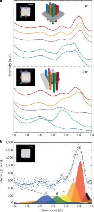
Energy filtered series for the 0° tilt (A)
and the −60° (B) STEM EELS spectrum images. The images are limited to the range of interest between 1 and 4 eV for LSPRs of the silver nanocube. The EELS signal in the images is integrated
over an energy window 0.2 eV wide. EXTENDED DATA FIGURE 3 DDA EELS SIMULATIONS OF A SILVER NANOCUBE. A, B, DDA EELS simulated as functions of position (as shown by the blue dotted line) for
a 100-nm silver cube with rounded corners in vacuum with dielectric function tabulated in ref. 33 (A), and for a 100-nm silver cube with rounded corners in vacuum with dielectric function
tabulated in ref. 42 (B). C, 2D tomographic reconstructions of the five spectral features identified in A and B (C1, C2, C3, E and F), made from a tilt series of simulated spectra in which
the cube is rotated about a cube axis perpendicular to the beam. The five spectral features correspond to lowest-energy (dipole) corner (C1), higher-energy corner (C2 and C3), edge (E) and
face (F) components. We note that the individual components C2 and C3 are not separated if the dielectric tabulation in ref. 42 is used, as in B. Each reconstruction is scaled independently.
There are significant negative values only in the reconstruction of the C3 component, which corresponds to a singly degenerate octupole mode. Tomographic reconstructions made from this tilt
series were obtained using SIRT41, implemented using the TOMO3D software package43. SUPPLEMENTARY INFORMATION 3D RENDERING OF THE LSPR COMPONENTS. This video shows a combined 3D rendering
of the LSPR components, as displayed in Fig. 3 of the manuscript. (MPG 16698 kb) 3D PERSPECTIVE VIEW OF EACH LSPR COMPONENT. This video shows a 3D perspective view of each LSPR component, in
respective energy order, leading to a combined 3D rendering, as displayed in Fig. 3 of the manuscript. (MPG 28893 kb) POWERPOINT SLIDES POWERPOINT SLIDE FOR FIG. 1 POWERPOINT SLIDE FOR FIG.
2 POWERPOINT SLIDE FOR FIG. 3 POWERPOINT SLIDE FOR FIG. 4 RIGHTS AND PERMISSIONS Reprints and permissions ABOUT THIS ARTICLE CITE THIS ARTICLE Nicoletti, O., de la Peña, F., Leary, R. _et
al._ Three-dimensional imaging of localized surface plasmon resonances of metal nanoparticles. _Nature_ 502, 80–84 (2013). https://doi.org/10.1038/nature12469 Download citation * Received:
22 April 2013 * Accepted: 15 July 2013 * Published: 02 October 2013 * Issue Date: 03 October 2013 * DOI: https://doi.org/10.1038/nature12469 SHARE THIS ARTICLE Anyone you share the following
link with will be able to read this content: Get shareable link Sorry, a shareable link is not currently available for this article. Copy to clipboard Provided by the Springer Nature
SharedIt content-sharing initiative