Light Emission from Plasmonic Nanostructures Enhanced with Fluorescent Nanodiamonds
- Select a language for the TTS:
- UK English Female
- UK English Male
- US English Female
- US English Male
- Australian Female
- Australian Male
- Language selected: (auto detect) - EN
Play all audios:

Download PDF Article Open access Published: 26 February 2018 Light Emission from Plasmonic Nanostructures Enhanced with Fluorescent Nanodiamonds Jingyi Zhao1 na1, Yuqing Cheng1 na1, Hongming
Shen1, Yuen Yung Hui2, Te Wen1, Huan-Cheng Chang2, Qihuang Gong1,3 & …Guowei Lu ORCID: orcid.org/0000-0003-0646-69711,3 Show authors Scientific Reports volume 8, Article number: 3605
(2018) Cite this article
6085 Accesses
Metrics details
Subjects NanoparticlesNanophotonics and plasmonicsSub-wavelength optics AbstractIn the surface-enhanced fluorescence (SEF) process, it is well known that the plasmonic nanostructure can enhance the light emission of fluorescent emitters. With the help of atomic force
microscopy, a hybrid system consisting of a fluorescent nanodiamond and a gold nanoparticle was assembled step-by-step for in situ optical measurements. We demonstrate that fluorescent
emitters can also enhance the light emission from gold nanoparticles which is judged through the intrinsic anti-Stokes emission owing to the nanostructures. The light emission intensity,
spectral shape, and lifetime of the hybrid system were dependent on the coupling configuration. The interaction between gold nanoparticles and fluorescent emitter was modelled based on the
concept of a quantised optical cavity by considering the nanodiamond and the nanoparticle as a two-level energy system and a nanoresonator, respectively. The theoretical calculations reveal
that the dielectric antenna effect can enhance the local field felt by the nanoparticle, which contributes more to the light emission enhancement of the nanoparticles rather than the
plasmonic coupling effect. The findings reveal that the SEF is a mutually enhancing process. This suggests the hybrid system should be considered as an entity to analyse and optimise
surface-enhanced spectroscopy.
Similar content being viewed by others Single-emitter super-resolved imaging of radiative decay rate enhancement in dielectric gap nanoantennas ArticleOpen access 02 January 2024 Sub-nanometre resolution in single-molecule photoluminescence imaging Article 10 August 2020 Achieving low-power single-wavelength-pair nanoscopy with NIR-II
continuous-wave laser for multi-chromatic probes Article Open access 23 May 2022 Introduction
Surface-enhanced fluorescence (SEF), enhancing the light emission of fluorescent emitters with metallic nanostructures, is the key to many promising applications1,2. By placing the emitters
in the vicinity of metallic nanostructures, the luminescence can be greatly enhanced because of the plasmon resonance effect. Metallic nanostructures support localised surface plasmon (LSP)
modes, which gives rise to a marked enhancement of local electromagnetic fields and results in a strongly modified local density of optical states3,4. Metallic nanostructures known as
optical antennas can be involved in the excitation and emission process of the emitters4,5,6,7,8. The metallic nanostructures enable the modification of the emission intensity, lifetime,
directivity, and spectral shape5,9,10,11,12,13,14. Basic theories of the SEF process have been developed15,16,17. In the conventional theory of SEF, there is a lack of attention on how the
fluorescent emitters influence the behaviour of the metallic nanostructures. Most of the previous SEF theories often assume that the light emission of the metal nanostructures is negligible
or stable5,18,19,20; this approximation allows us to mostly understand SEF.
The SEF background was often simply subtracted as a stable signal when calculating the SEF enhancement factor. A deviation of the enhancement factor is inevitable if the emission from the
metal nanostructures changes owing to the presence of emitters. Furthermore, this approximation often hinders us from fully and accurately understanding the SEF spectral shape4,21,22,23.
Intrinsic light emission from metal nanostructures has been demonstrated as a complementary property to absorption and scattering24,25,26. Although the mechanism is still under debate, a
solid consensus is that the metal nanostructures can radiate at their LSP resonances after photon or electron excitation27,28. Such light emission (here, it is called photoluminescence (PL))
from the nano-metal can be correlated with the broad “background continuum” in the SEF process29,30.
The interactions between fluorescent emitters and metallic nanostructures has been extensively investigated experimentally31,32. Despite much progress, the changes in the light emission of
the plasmonic nanostructures influenced by the fluorescent emitters have often been ignored in previous studies. Access to experimental analysis of light emission of plasmonic nanostructures
during the SEF process has also proven elusive for a long time. This problem has not been addressed in many recent studies, probably because of the experimental challenge of isolating the
intrinsic emission signal of the metallic nanostructures; the PL of the metallic nanoparticles is usually broad and weak, and which often overlaps with the light emission of fluorescent
emitters. As a result, it is nontrivial to observe the changes of the intrinsic emission from the metallic nanoparticles during the SEF process. Here, we found that the difference in the
emission between the metal nanostructures and the fluorescent emitters can be distinguished through the anti-Stokes emission, which enabled us to circumvent previous experimental challenges.
Specifically, we performed optical measurements at the single particle level to avoid inhomogeneous average-broadened spectra. The light emission from the same gold nanoparticles and
nanodiamonds were recorded in situ and compared on the same substrate.
In this study, we experimentally demonstrate that the light emission from the gold nanostructures is not a constant signal and is enhanced when couple with fluorescent emitters. Not only can
the gold nanostructure enhance the light emission of the fluorescent emitters, as expected, but the emitters can also increase the light emission from the gold nanostructures. With the help
of an atomic force microscope (AFM), a single gold nanoparticle was manipulated step-by-step to approach a single fluorescent nanodiamond (FND) on a glass coverslip. The emission spectra
excited with a continuous-wave laser were recorded in situ and compared before and after coupling. We found that the emission from the gold nanoparticle can be distinguished from the SEF
hybrid spectra through its characteristic anti-Stokes emission because the FND does not present any anti-Stokes emission. According to the experimental results, the light emission from the
FND and gold nanoparticle both enhanced after coupling. The theoretical calculations reveal that both the plasmonic coupling effect and the dielectric nanoantenna effect contribute to the
enhancement of light emission from the nanoparticles. Specifically, the dielectric nanoantenna effect dominates the emission enhancements from the plasmonic nanostructures, as confirmed by
correlating the theoretical and experimental results.
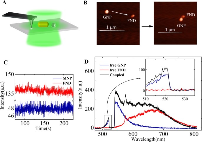
Experimental ResultsWe used gold nanoparticles (GNP) and gold nanorods (GNR) to couple with FND for the SEF, as shown in Fig. 1A,B. The representative PL spectrum of a free GNP (Fig. 1D) shows well-defined
emission bands rather than the broad continuum band usually associated with a rough thin film, which is a benefit for the analysis of the surface plasmon effect in SEF33. The emission
spectra of the FNDs and GNPs were both very stable (nonbleaching and nonblinking), as shown in Fig. 1C34, which is important for in situ comparison before and after coupling. Specifically,
as illustrated in Fig. 1D, the emission spectra of the FNDs dominate the range of Stokes components, but do not have any signal in the anti-Stokes range. In contrast, the emission spectra of
the GNPs have an obvious anti-Stokes band that decays exponentially as a function of photon energy. The one-photon luminescent anti-Stokes emission of metallic nanostructures has been
demonstrated in different metal nanostructures, although the mechanism is still the subject of much debate27,29,30,33,35. Interestingly, this anti-Stokes component allows us to differentiate
the intrinsic light emission of the GNPs from the SEF hybrid spectra. As shown in the inset of Fig. 1D, the intensity of the anti-Stokes emission from the coupling system increases in
comparison with that before coupling. The enhancement factor can be defined as I c /I uc , in which I c andI uc are the maximum of the PL spectra before and after coupling, respectively. For
the anti-Stokes emission, the intensity increased over 40% at a wavelength of 520 nm. There have been numerous reports in the literature about the coupling phenomena of nanodiamonds and
plasmonic structures31,32, but the anti-Stokes emission has been seldom discussed. Because the anti-Stokes emission is solely owing to the GNP in the present system, this implies that the
FND enhances the light emission from the GNPs. The same measurements were performed for the GNR and FND coupling system, as shown in Fig. S1A. The intensity of the spectrum of the coupled
GNR and FND system in the Stokes range increased compared to that before coupling, and even compared to the sum of the intensities in the free GNR and FND spectra. The spectral peak of the
SEF undergoes a redshift compared to that of the free FND spectrum. For the anti-Stokes component, the intensity of the SEF was also enhanced compared with that of the GNR before coupling.
Moreover, the lifetime of the SEF system was also investigated, as shown in Fig. S1B. The lifetime of the free GNR was less than 1 ps, which was faster than the instrument response. The
lifetime curves of the free FND can be fitted with two exponential components. Consequently, the lifetime curve of the GNR–FND coupling system contains three exponential components, the
coupling between the GNR and FND results in a shorter lifetime of one component owing to the FND. The spectra of the GNRs, FNDs, and their hybrids showed excitation polarisation dependent
characteristics as indicated in the supplemental materials. To demonstrate the enhancement of anti-Stokes emission definitely, we measured overall PL spectra of a nanorod before and after
coupling with a nanodiamond under different excitation polarisations. Then, we choose two maximum spectra respectively from two series of the spectra (before coupling and after coupling) for
comparison, hence, the excitation polarisation effect was excluded indirectly. We also excluded the influences of the excitation power fluctuation and the uncertainty owing to the optical
path drifts, which is depicted in Figs S2 and 3. Hence, the SEF phenomena is a mutually enhancing process owing to the interaction between the fluorescent emitters and the gold
nanostructures. To calculate the SEF enhancement factor and understand the SEF spectral shape, the change of the light emission from the metal nanostructures must be considered.
Figure 1Scheme of nano-manipulation and representative in situ optical measurements. (A) Scheme of the AFM manipulation method, (B) representative AFM images during the assembly process. (C) Time
trace of the PL intensity of a gold nanoparticle and a nanodiamond. (D) The PL spectra of a free GNP (dark blue) and a free FND (red) before coupling and the SEF spectrum (black) after
coupling. The inset shows a magnification of the area showing the anti-Stokes component.
Full size imageFurthermore, as shown in Figs 1D and 2A, the SEF spectral shape in the Stokes range is totally different from that of free FND or free GNP. It is well-known that the plasmonic nanostructures
enable the modification of the emission spectral shape4. Ringler et al. have developed an empirical formula to correlate the scattering and luminescence spectral shape of GNP dimers and dye
molecules systems11. Based on their work, we show that the emission from the GNP should be taken into account to fully understand the SEF spectra. We supposed that a SEF spectrum S SEF
contains three components: two direct emissions S GNR , S FND are from the GNR and FND separately, and one indirect emission (S gc ) is owing to the optical antenna coupling effect, i.e., S
SEF = S GNR + S FND + S gc . The direct emission S GNR , based on the following quantum model, is inelastic decay radiation of the plasmon resonance of the GNR. The indirect emission S gc
can be understood as a process of elastic radiation by the antenna after coupling energy from excited states of the fluorescence emitters11. We assumed that the coupling rate Γ g between the
FND and the GNR is less than the internal phonon decay rate of the FND and the frequency-dependent Γ g is strongly related to the LSP resonance, i.e., \({{\rm{\Gamma }}}_{g} \sim
\,{\hat{{\rm{S}}}}_{SCA}\) (the hybrid scattering spectrum)6,12,36. Then, we obtained the three components by fitting the SEF spectrum using
\({{\rm{S}}}_{SEF}={I}_{1}{{\rm{S}}}_{GNR0}+{I}_{2}{{\rm{S}}}_{FND0}+{g}_{c}{\hat{{\rm{S}}}}_{SCA}{\hat{{\rm{S}}}}_{FND0}\), in which the terms with a 0 subscript are the spectra before
coupling, \(\hat{{\rm{S}}}\) is the normalised spectrum, and I1,I2,and g c are the fitting constant parameters. Figure 2A shows the experimental spectra of SGNR0 and SFND0 before coupling,
and the S SEF increasing in both the Stokes and anti-Stokes region after coupling. The fitting results are summarised in Fig. 2B. The direct emission from the GNR and the FND increased by
40% and 140% respectively, and the indirect emission photons by the antenna effect was approximately 43% of the whole SEF spectra. Therefore, the emission from the metallic nanostructures
can no longer be omitted or assumed to be constant when calculating the SEF enhancement factor. All three components determine the final SEF spectral shape. However, the integrated intensity
of the anti-Stokes emission of the GNR is relatively small; thus, the total modification of the emitted power differs only by a few percent, which indicates that the emission from the metal
is necessary to accurately and fully understand the SEF spectral shape. The SEF spectrum of the GNP-FND system shown in Fig. S4 was analysed in the same way as that of the GNR-FND system in
Fig. 2B. The SEF spectra was modified by the GNP antenna effect, and the direct emission from the GNP dominated the SEF spectra near the excitation due to the GNP’s LSP resonance
band.
Figure 2A complete analysis of the three components of the SEF spectrum and coupling-configuration-dependent SEF spectra. (A) The PL spectra of a free GNR (black) and a free FND (red) before
coupling, the SEF spectrum (blue) and scattering spectrum (green) after the coupling of GNR and FND. (B) Fitting spectra for I1SGNR0, I2SFND0, and
\({g}_{c}{\hat{{\rm{S}}}}_{SCA}{\hat{{\rm{S}}}}_{FND0}\). (C) Configuration-dependent SEF spectra of the hybrid. The spectra of a free GNR (blue) and a free FND (red) before coupling, and
three SEF spectra of three different configurations, as indicated by the AFM and schematic images. The inset shows the lifetime curves of the config. 1 and config. 3 configurations for
comparison.
Full size imageIn addition, we showed that the SEF spectral shape, intensity, and lifetime vary for different coupling configurations between the GNR and FND6,37. The distance between the FND and GNR
before coupling was several micrometres, which was enough to isolate them by optics. However, the exact distance between the FND and GNR after coupling could not be determined, with an error
of approximately 10 nm. Figure 2C shows the SEF spectra of a GNR and FND hybrid system with a different configuration obtained through AFM nanomanipulations. It is well known that the
coupling rate Γ g (i.e., being related with the Purcell factor) is dependent on separation and orientation of the emitter with respect to the antenna36,38. Then, the indirect emission S gc
intensity is dependent on the coupling strength, i.e., the coupling configuration. The Purcell factor distribution can be similar to the near-field distribution of the GNR36. When the FND
was closer to the GNR end, the spectral maximum increased and redshifted more, and the lifetime was shorter. Because the coupling strength increased, the indirect emission component
increased, which significantly modified the spectral shape. Another factor is that the FND as a dielectric particle would increase the local effective index felt by the GNR, resulting in a
redshift of the LSP resonance as well as the SEF spectra39. We noted that the fluorescence of nanodiamond is excitation polarisation dependent, the polarisation contributions can be excluded
roughly by comparing the spectra of nanodiamond of the same excitation polarisation before and after coupling because the nanodiamonds were always fixed in the experiments. Here, we
demonstrate that the SEF process is strongly dependent on the coupling configurations.
Theoretical Model and DiscussionsTo understand the physical origin of the enhancement of direct emission from the gold nanoparticles, we give a theoretical description of the interactions based on the concept of a quantised
optical cavity, as shown in Fig. 3A. The GNR and FND hybrid was modelled as an entity consisting of a two-level atom and a nanoresonator cavity. We considered a FND interacting with a GNR
separated by a distance. There is no direct electron tunnelling between the GNR and FND. The coupling mechanism is owing to dipole–dipole interaction. The artificial hybrid system is excited
with a quasi-continuous-wave laser beam with frequency ω ex polarised along the system axis. Considering the GNR as a plasmonic resonator, the Hamiltonian of the LSP resonator with mode a
at ω c is described as H c = ω c a†a. Considering the FND as a two-energy-level atom system, the Hamiltonian is written as \({H}_{m}={\omega }_{g}\,|g\rangle \langle g|+{\omega
}_{e}|e\rangle \langle e|\), in which the energy difference between \(|g\rangle \) and \(|e\rangle \) is ω em = ω e −ω g . Specifically, we define σ− = \(|g\rangle \)\(\langle e|\),σ+ =
\(|e\rangle \)\(\langle g|\) as the transition operators. Therefore, the free Hamiltonian of the whole system without any interaction is written as: H0 = H c + H m = ω c a†a + ω g
\(|g\rangle \) \(\langle g|\) + ω e \(|e\rangle \) \(\langle e|\). The interaction Hamiltonian is described as: \({H}_{I}=g({a}^{\dagger }{\sigma }_{-}+a{\sigma }_{+})+{\mu
}_{1}{E}_{1}({a}^{\dagger }{e}^{-i{\omega }_{ex}t}+a{e}^{i{\omega }_{ex}t})+{\mu }_{2}{E}_{2}({\sigma }_{+}{e}^{-i{\omega }_{ex}t}+{\sigma }_{-}{e}^{i{\omega }_{ex}t})\), in which the first
term demonstrates that the LSP mode couples with states \(|g\rangle \) and \(|e\rangle \), and g is the coupling constant. The second and the third terms show that the excitation
electromagnetic field couples with the LSP mode and states \(|g\rangle \) and \(|e\rangle \), respectively. μ1 and μ2 are the corresponding coupling constants. E1 and E2 are the respective
localised electromagnetic field amplitudes that the GNR and the atom feel. Hence, the Hamiltonian of this system is given by H = H0 + H I . The dynamics of these modes can be solved by the
equations \(\dot{a}=i[H,a]-\kappa a=(-i{\omega }_{c}-\kappa )a-ig{\sigma }_{-}-i{\mu }_{1}{E}_{1}{e}^{-i{\omega }_{ex}t}\) and \({\dot{\sigma }}_{-}=i[H,{\sigma }_{-}]\) \(-\,\gamma {\sigma
}_{-}=(-i{\omega }_{em}-\gamma ){\sigma }_{-}+iga{\sigma }_{z}+i{\mu }_{2}{E}_{2}{\sigma }_{z}{e}^{-i{\omega }_{ex}t}\), where κ and γ are the total decay of the cavity and the atom,
respectively. σ z = \(|e\rangle \) \(\langle e|\) − \(|g\rangle \) \(\langle g|\), which represents the difference between the level occupation numbers of states \(|e\rangle \) and
\(|g\rangle \). Details of the solution are shown in the supplemental materials. We assume that \(g\ll k-\gamma \) because the SEF process is a weak coupling system37,40,41,42. This is
supported by the experimental data
, whereby the frequency of the zero-phonon line of the FND does not shift after coupling with the GNR.
Figure 3Scheme of the interaction between a FND and a GNR and theoretical analysis of the influence of the dipole coupling effect and local field of the dielectric particle. Simulated spectra of a
resonator (black dash) and an atom (black dot) for g = 0, and corresponding spectra (solid curves) after coupling for g = 10 meV (A) the applied electromagnetic field E induces polarisations
that causes dipole–dipole coupling Γ g between the resonator and the atom and light emission \({\Gamma }_{r}^{LSP}\) from the GNR including both elastic and inelastic radiation processes.
(B) Local field felt by the GNR for E1 = E0, i.e., without the dielectric nanoantenna effect (C) for E1 = 1.1E0 owing to the induced field of the polarised FND. The inset in (B) presents an
enlarged view of the enhanced emission of the resonator.
Full size imageFor g = 0, which suggests no coupling between the GNR and FND, we obtained two Lorentz-shape spectra, a narrow peak for the atom and a broad band for the nanoresonator. When the coupling
effect occurs (e.g., g = 10 meV) and the atom undergoes an enhanced excitation field owing to the plasmonic near field of the nanorod, then it is set as E2 = 1.5E0. At first, we assume that
the presence of the nanodiamond does not influence the local field felt by the GNR, i.e., E1 = E0. Then, the emission from the nanoresonator increases slightly (less than 10%), as shown in
Fig. 3B. This can be explained that total dipole moment of the NV cements in the FND is smaller than that of the GNP which resulting in low energy transfer rate, and that the inelastic
plasmon radiation efficiency of the GNP is also very low. Hence the plasmon emission intensity due to the energy coupling from the FND should be low leading to small increasing. However, the
enhancement of direct emission from the GNP can be over 40% in the above experiment. Then, the coupling effect cannot fully explain the total change. Therefore, we increased E1 and set it
asE 1 = 1.1E0, i.e., the presence of the FND increases the local field felt by the GNR. We obtained a considerable enhancement of the nanoresonator emission, as shown in Fig. 3C. This
implied that the induced field produced by the polarisation of the FND increased the local field intensity felt by the GNR. A dielectric nanoparticle (DNP) as a non-plasmonic nanoantenna
should be the main factor for the enhanced emission from a gold nanoparticle43. More details about the theoretical model is discussed in the supplemental materials.
The dielectric nanoantenna effect is supported by the finite-difference time-domain (FDTD) numerical calculations shown in Fig. 4A–C. The DNP presents an enhanced local field that can
increase the EM field felt by the GNR. Hence, in the SEF process, the light emission from the atom and nanoresonator both increase simultaneously. The plasmonic antenna effect and the
dielectric nanoantenna effect contribute to this mutual enhancement. The influence of radiative directivity on the light collection efficient was negligible. As shown in Fig. 4D, the
emission patterns of a GNR was simulated using the FDTD method. The patterns were almost the same before and after coupling with a DNP. The presence of DNP (i.e., increasing of the effective
index for upper half-space) decreases the electromagnetic flux toward the glass substrate by less than 1% compared with that of the free GNR. So far, the theoretical results reveal that the
plasmonic coupling effect can significantly enhance light emission from the emitter; it also results in a slight enhanced emission from the GNPs through the plasmon decay radiation. The
enhancement of local field felt by the GNP should dominate the enhanced emission from the gold nanoparticles.
Figure 4Field distributions (X–Z plane) of (A) a GNR with a DNP, (B) only a DNP, and (C) only a GNR. (B) The DNP with a diameter of 40 nm and refractive index of 2.3 in the FDTD calculations; the
averaged field felt by the GNR (white dot range) is higher than the external excitation filed. (C) Near-field EM distribution around a GNR (60 nm×120 nm) at a wavelength of 532 nm. The white
dot range shows the region of the DNP. (D) Ratio of emission flux from a GNR toward a glass substrate to total flux with (black dot) or without (green solid) the DNP as a function of
wavelength. The inset in (D) shows representative emission patterns toward a glass substrate or air as indicated at a wavelength of 680 nm.
Full size imageConclusionIn summary, we have demonstrated experimentally and theoretically that gold nanoparticles and fluorescent nanodiamonds mutually enhance their light emission when they are coupled for the SEF
process. The light emission of the metal nanostructures should be considered for quantifying the SEF enhancement factor and for understanding the SEF spectral shape. These findings
contribute to full and deep understanding of the SEF process. The SEF background arising from the metallic nanostructure is not noise or a constant background. The light emission from the
metallic nanostructure is another indicator of the interaction strength between the metallic nanostructure and the emitter. The observation introduces a new perspective in the field of
surface-enhanced spectroscopy to fully understand SEF process. For instance, the mutual interaction is also effective in surface-enhanced Raman scattering44. The coupling system for
surface-enhanced spectroscopy should be considered as a hybrid entity to analyse and optimise.
Materials and MethodsIn our experiments, FNDs with a nominal size of 35 nm were prepared by radiation damage of type-Ib diamond powders containing several nitrogen vacancy centers45. The GNPs with diameter of
approximately 80 nm and GNRs with size of approximately 60 nm × 100 nm were synthesised through a seed-mediated wet chemical method46. A dilute aqueous solution containing both the FNDs and
GNRs was cast onto a silane-functionalised glass coverslip. Then, the nanoparticles were immobilised, with an average spacing of several micrometres for single particle-level investigations.
We characterised the system optically with a micro-spectroscopy system using an inverted optical microscope combined with an AFM, as shown schematically in Fig. 1A. A continuous-wave laser
at a wavelength of 532 nm passing through an objective lens was used to excite the samples, and the fluorescent emission was recorded through the same objective lens. The light emission was
recorded in situ before and after coupling. Moreover, the fluorescence signal from the same particles could be switched to an avalanche photodiode, the lifetime and time trace were analysed
with a TCSPC module (PicoHarp 300, PicoQuant). In this case, a picosecond laser diode operating at a wavelength of 480 nm with a repeat rate of 10 MHz was implemented for the fluorescence
lifetime measurements. By AFM nanomanipulation, the GNPs were moved to approach the FNDs step-by-step, as shown in Fig. 1B31,47,48,49,50. In addition, the scattering spectra of the same GNPs
was obtained in situ by the white light total internal reflection dark field method. Furthermore, the three-dimensional FDTD method was employed to simulate the emission flux, emission
patterns, and electromagnetic field distribution of the nanostructures51. The calculations of the far-field radiation pattern were based on the near-field-to-far-field (NFTFF) transformation
method. For the NFTFF calculations, we chose a large transformation plane (2 μm × 2 μm) placed 20 nm beneath the air/glass interface to collect most of the flux directed to the substrate.
In the simulations, the mesh size was 1 nm to match the memory resources and computation time. The optical dielectric function of gold was modelled using the Drude–Lorentz dispersion
model52. The refractive indices of the material were set to 1.0 for air and 1.49 for the glass substrate.
References Moskovits, M. Surface-enhanced spectroscopy. Rev. Mod. Phys. 57, 783 (1985).
Article ADS CAS Google Scholar
Fort, E. & Grésillon, S. Surface enhanced fluorescence. J. Phys. D: Appl. Phys. 41, 013001 (2008).
Article ADS Google Scholar
Acuna, G. P. et al. Fluorescence enhancement at docking sites of DNA-Directed self-assembled nanoantennas. Science 338, 506 (2012).
Article ADS CAS PubMed Google Scholar
Pelton, M. Modified spontaneous emission in nanophotonic structures. Nat. Photon. 9, 427 (2015).
Article ADS CAS Google Scholar
Anger, P., Bharadwaj, P. & Novotny, L. Enhancement and quenching of single-molecule fluorescence. Phys. Rev. Lett. 96, 113002 (2006).
Article ADS PubMed Google Scholar
Vecchi, G., Giannini, V. & Gomez Rivas, J. Shaping the fluorescent emission by lattice resonances in plasmonic crystals of nanoantennas. Phys. Rev. Lett. 102, 146807 (2009).
Article ADS CAS PubMed Google Scholar
Gerber, S. et al. Tailoring light emission properties of fluorophores by coupling to resonance-tuned metallic nanostructures. Phys. Rev. B 75, 073404 (2007).
Article ADS Google Scholar
Akselrod, G. M. et al. Probing the mechanisms of large Purcell enhancement in plasmonic nanoantennas. Nat. Photon. 8, 835 (2014).
Article ADS CAS Google Scholar
de Leon, N. P. et al. Tailoring light-matter interaction with a nanoscale plasmon resonator. Phys. Rev. Lett. 108, 226803 (2012).
Article ADS PubMed Google Scholar
Galloway, C. M., Etchegoin, P. G. & Le, R. E. C. Ultrafast nonradiative decay rates on metallic surfaces by comparing surface-enhanced Raman and fluorescence signals of single molecules.
Phys. Rev. Lett. 103, 063003 (2009).
Article ADS CAS PubMed Google Scholar
Ringler, M. et al. Shaping emission spectra of fluorescent molecules with single plasmonic nanoresonators. Phys. Rev. Lett. 100, 203002 (2008).
Article ADS CAS PubMed Google Scholar
Karaveli, S. & Zia, R. Spectral tuning by selective enhancement of electric and magnetic dipole emission. Phys. Rev. Lett. 106, 193004 (2011).
Article ADS PubMed Google Scholar
Russell, K. J., Liu, T.-L., Cui, S. & Hu, E. L. Large spontaneous emission enhancement in plasmonic nanocavities. Nat. Photon. 6, 459 (2012).
Article ADS CAS Google Scholar
Pompa, P. P. et al. Metal-enhanced fluorescence of colloidal nanocrystals with nanoscale control. Nat. Nanotech. 1, 126 (2006).
Article ADS CAS Google Scholar
Andrew, P. & Barnes, W. L. Energy transfer across a metal film mediated by surface plasmon polaritons. Science 306, 1002 (2004).
Article ADS CAS PubMed Google Scholar
Sun, G., Khurgin, J. B. & Soref, R. A. Practical enhancement of photoluminescence by metal nanoparticles. Appl. Phys. Lett. 94, 101103 (2009).
Article ADS Google Scholar
Schlather, A. E., Large, N., Urban, A. S., Nordlander, P. & Halas, N. J. Near-field mediated plexcitonic coupling and giant Rabi splitting in individual metallic dimers. Nano Lett. 13, 3281
(2013).
Article ADS CAS PubMed Google Scholar
Yang, J., Perrin, M. & Lalanne, P. Analytical formalism for the interaction of wwo-level quantum systems with metal nanoresonators. Phys. Rev. X 5, 021008 (2015).
Google Scholar
Kinkhabwala, A. et al. Large single-molecule fluorescence enhancements produced by a bowtie nanoantenna. Nat. Photon. 3, 654 (2009).
Article ADS CAS Google Scholar
Johansson, P., Xu, H. & Käll, M. Surface-enhanced Raman scattering and fluorescence near metal nanoparticles. Phys. Rev. B 72, 035427 (2005).
Article ADS Google Scholar
Darby, B. L., Auguié, B., Meyer, M., Pantoja, A. E. & Le Ru, E. C. Modified optical absorption of molecules on metallic nanoparticles at sub-monolayer coverage. Nat. Photon. 10, 40 (2015).
Article ADS Google Scholar
Dong, Z. C. et al. Generation of molecular hot electroluminescence by resonant nanocavity plasmons. Nat. Photon. 4, 50 (2010).
Article ADS CAS Google Scholar
Cho, C. H., Aspetti, C. O., Park, J. & Agarwal, R. Silicon coupled with plasmon nanocavities generates bright visible hot luminescence. Nat. Photon. 7, 285 (2013).
Article ADS CAS Google Scholar
Yorulmaz., M., Khatua, S., Zijlstra, P., Gaiduk, A. & Orrit, M. Luminescence quantum yield of single gold nanorods. Nano Lett. 12, 4385 (2012).
Article ADS CAS PubMed Google Scholar
Cheng, Y. et al. Luminescence quantum yields of gold nanoparticles varying with excitation wavelengths. Nanoscale 8, 2188 (2016).
Article ADS CAS PubMed Google Scholar
Carles, R. et al. Plasmon-resonant Raman spectroscopy in metallic nanoparticles: Surface-enhanced scattering by electronic excitations. Phys. Rev. B 92, 174302 (2015).
Article ADS Google Scholar
Haug, T., Klemm, P., Bange, S. & Lupton, J. M. Hot-electron intraband luminescence from single hot spots in noble-metal nanoparticle films. Phys. Rev. Lett. 115, 067403 (2015).
Article ADS PubMed Google Scholar
Dulkeith, E. et al. Plasmon emission in photoexcited gold nanoparticles. Phys. Rev. B 70, 205424 (2004).
Article ADS Google Scholar
Hugall, J. T. & Baumberg, J. J. Demonstrating photoluminescence from Au is electronic inelastic light scattering of a plasmonic metal. Nano Lett. 15, 2600 (2015).
Article ADS CAS PubMed PubMed Central Google Scholar
Huang, J., Wang, W., Murphy, C. J. & Cahill, D. G. Resonant secondary light emission from plasmonic Au nanostructures at high electron temperatures created by pulsed-laser excitation. PNAS
111, 906 (2014).
Article ADS CAS PubMed PubMed Central Google Scholar
Schietinger, S., Barth, M., Alchele, T. & Benson, O. Plasmon-enhanced single photon emission from a nanoassembled metal-diamond hybrid structure at room temperature. Nano Lett. 9, 1694
(2009).
Article ADS CAS PubMed Google Scholar
Hui, Y. Y. et al. Tip-enhanced sub-diffraction fluorescence imaging of nitrogen-vacancy centers in nanodiamonds. Appl. Phys. Lett. 102, 013102 (2013).
Article ADS Google Scholar
He, Y. et al. Surface enhanced anti-Stokes one-photon luminescence from single gold nanorods. Nanoscale 7, 577 (2015).
Article ADS CAS PubMed Google Scholar
Fu, C. C. et al. Characterization and application of single fluorescent nanodiamonds as cellular biomarkers. PNAS 104, 727 (2007).
Article ADS CAS PubMed PubMed Central Google Scholar
Neupane, B., Zhao, L. Y. & Wang, G. F. Up-conversion luminescence of gold nanospheres when excited at nonsurface plasmon resonance wavelength by a continuous wave laser. Nano Lett. 13, 4087
(2013).
Article ADS CAS PubMed Google Scholar
Lu, G. et al. Single-molecule spontaneous emission in the vicinity of an individual gold nanorod. J. Phys. Chem. C 115, 15822 (2011).
Article CAS Google Scholar
Ming, T., Chen, H., Jiang, R., Li, Q. & Wang, J. Plasmon-controlled fluorescence: beyond the intensity enhancement. J. Phys. Chem. Lett. 3, 191 (2012).
Article CAS Google Scholar
Hartling, T., Reichenbach, P. & Eng, L. M. Near-field coupling of a single fluorescent molecule and a spherical gold nanoparticle. Opt. Express 15, 12806 (2007).
Article ADS CAS PubMed Google Scholar
Alaverdyan, Y. et al. Spectral tunability of a plasmonic antenna with a dielectric nanocrystal. Opt. Express 19, 18175 (2011).
Article ADS CAS PubMed Google Scholar
Ni, W., Ambjornsson, T. S., Apell, P., Chen, H. & Wang, J. Observing plasmonic-molecular resonance coupling on single gold nanorods. Nano Lett. 10, 77 (2010).
Article ADS CAS PubMed Google Scholar
Zhang, W., Govorov, A. O. & Bryant, G. W. Semiconductor-metal nanoparticle molecules: Hybrid excitons and the nonlinear Fano effect. Phys. Rev. Lett. 97, 146804 (2006).
Article ADS PubMed Google Scholar
Ridolfo, A., Di Stefano, O., Fina, N., Saija, R. & Savasta, S. Quantum plasmonics with quantum dot-metal nanoparticle molecules: influence of the Fano effect on photon statistics. Phys. Rev.
Lett. 105, 263601 (2010).
Article ADS CAS PubMed Google Scholar
Caldarola, M. et al. Non-plasmonic nanoantennas for surface enhanced spectroscopies with ultra-low heat conversion. Nat. Commun. 6, 7915 (2015).
Article CAS PubMed PubMed Central Google Scholar
Cheng, Y. et al. Enhanced light emission from plasmonic nanostructures by molecules. J. Phys. Chem. C 121, 23626 (2017).
Article CAS Google Scholar
Chang, Y. R. et al. Mass production and dynamic imaging of fluorescent nanodiamonds. Nat. Nanotech. 3, 284 (2008).
Article CAS Google Scholar
Zhang, T. et al. Single Bipyramid plasmonic antenna orientation determined by direct photoluminescence pattern imaging. Adv. Opt. Mater. 1, 335 (2013).
Article ADS Google Scholar
Shen, H. et al. Directional fluorescence emission from a compact plasmonic-diamond hybrid nanostructure. Laser & Photon. Rev. 10, 647 (2016).
Article CAS Google Scholar
Junno, T., Deppert, K., Montelius, L. & Samuelson, L. Controlled manipulation of nanoparticles with an atomic-force microscope. Appl. Phys. Lett. 66, 3627 (1995).
Article ADS CAS Google Scholar
Kim, S., Shafiei, F., Ratchford, D. & Li, X. Controlled AFM manipulation of small nanoparticles and assembly of hybrid nanostructures. Nanotech. 22, 115301 (2011).
Article ADS Google Scholar
Benson, O. Assembly of hybrid photonic architectures from nanophotonic constituents. Nature 480, 193 (2011).
Article ADS CAS PubMed Google Scholar
Oskooi, A. F. et al. Benson O. Assembly of hybrid photonic architectures from nanophotonic constituents. Comp. Phys. Commun. 181, 687 (2010).
Article ADS CAS Google Scholar
Johnson, P. B. & Christy, R. W. Optical Constants of the noble metals. Phys. Rev. B 6, 4370 (1972).
Article ADS CAS Google Scholar
Download references
AcknowledgementsThis work was supported by the National Key Basic Research Program of China (grant no. 2013CB328703) and the National Natural Science Foundation of China (grant nos 61422502, 11374026,
61521004, 11527901).
Author informationAuthor notesJingyi Zhao and Yuqing Cheng contributed equally to this work.
Authors and Affiliations State Key Laboratory for Mesoscopic Physics & Collaborative Innovation Center of Quantum Matter, Department of Physics, Peking University, Beijing, 100871, China
Jingyi Zhao, Yuqing Cheng, Hongming Shen, Te Wen, Qihuang Gong & Guowei Lu
Institute of Atomic and Molecular Sciences, Academia Sinica, Taipei, 104 Taiwan, China
Yuen Yung Hui & Huan-Cheng Chang
Collaborative Innovation Center of Extreme Optics, Shanxi University, Taiyuan, Shanxi, 030006, China
Qihuang Gong & Guowei Lu
AuthorsJingyi ZhaoView author publications You can also search for this author inPubMed Google Scholar
Yuqing ChengView author publications You can also search for this author inPubMed Google Scholar
Hongming ShenView author publications You can also search for this author inPubMed Google Scholar
Yuen Yung HuiView author publications You can also search for this author inPubMed Google Scholar
Te WenView author publications You can also search for this author inPubMed Google Scholar
Huan-Cheng ChangView author publications You can also search for this author inPubMed Google Scholar
Qihuang GongView author publications You can also search for this author inPubMed Google Scholar
Guowei LuView author publications You can also search for this author inPubMed Google Scholar
ContributionsG.W.L. designed the original ideas presented in this work and built the experimental setup. J.Y.Z. and H.M.S. carried out the optical measurements. Y.Y.H. and H.C.C. prepared the
nanodiamonds, and T.W. prepared the gold nanoparticles. Y.Q.C. developed and performed the theoretical calculations. The manuscript was jointly written by G.W.L., J.Y.Z., and Q.H.G. All
authors discussed the results and the manuscript.
Corresponding author Correspondence to Guowei Lu.
Ethics declarations Competing InterestsThe authors declare no competing interests.
Additional informationPublisher's note: Springer Nature remains neutral with regard to jurisdictional claims in published maps and institutional affiliations.
Electronic supplementary materialSupplementarymaterialsRights and permissions
Open Access This article is licensed under a Creative Commons Attribution 4.0 International License, which permits use, sharing, adaptation, distribution and reproduction in any medium or
format, as long as you give appropriate credit to the original author(s) and the source, provide a link to the Creative Commons license, and indicate if changes were made. The images or
other third party material in this article are included in the article’s Creative Commons license, unless indicated otherwise in a credit line to the material. If material is not included in
the article’s Creative Commons license and your intended use is not permitted by statutory regulation or exceeds the permitted use, you will need to obtain permission directly from the
copyright holder. To view a copy of this license, visit http://creativecommons.org/licenses/by/4.0/.
Reprints and permissions
About this articleCite this article Zhao, J., Cheng, Y., Shen, H. et al. Light Emission from Plasmonic Nanostructures Enhanced with Fluorescent Nanodiamonds. Sci Rep 8, 3605 (2018).
https://doi.org/10.1038/s41598-018-22019-z
Download citation
Received: 07 November 2017
Accepted: 15 February 2018
Published: 26 February 2018
DOI: https://doi.org/10.1038/s41598-018-22019-z
Share this article Anyone you share the following link with will be able to read this content:
Get shareable link Sorry, a shareable link is not currently available for this article.
Copy to clipboard Provided by the Springer Nature SharedIt content-sharing initiative