Premature termination codon readthrough in drosophila varies in a developmental and tissue-specific manner
- Select a language for the TTS:
- UK English Female
- UK English Male
- US English Female
- US English Male
- Australian Female
- Australian Male
- Language selected: (auto detect) - EN
Play all audios:

ABSTRACT Despite their essential function in terminating translation, readthrough of stop codons occurs more frequently than previously supposed. However, little is known about the
regulation of stop codon readthrough by anatomical site and over the life cycle of animals. Here, we developed a set of reporters to measure readthrough in _Drosophila melanogaster_. A
focused RNAi screen in whole animals identified _upf1_ as a mediator of readthrough, suggesting that the stop codons in the reporters were recognized as premature termination codons (PTCs).
We found readthrough rates of PTCs varied significantly throughout the life cycle of flies, being highest in older adult flies. Furthermore, readthrough rates varied dramatically by tissue
and, intriguingly, were highest in fly brains, specifically neurons and not glia. This was not due to differences in reporter abundance or nonsense-mediated mRNA decay (NMD) surveillance
between these tissues. Readthrough rates also varied within neurons, with cholinergic neurons having highest readthrough compared with lowest readthrough rates in dopaminergic neurons.
Overall, our data reveal temporal and spatial variation of PTC-mediated readthrough in animals, and suggest that readthrough may be a potential rescue mechanism for PTC-harboring transcripts
when the NMD surveillance pathway is inhibited. SIMILAR CONTENT BEING VIEWED BY OTHERS DIVERSE CELL-SPECIFIC PATTERNS OF ALTERNATIVE POLYADENYLATION IN _DROSOPHILA_ Article Open access 13
September 2022 AN IN VIVO STRATEGY FOR KNOCKDOWN OF CIRCULAR RNAS Article Open access 11 August 2020 ORB2 ENABLES RARE-CODON-ENRICHED MRNA EXPRESSION DURING _DROSOPHILA_ NEURON
DIFFERENTIATION Article Open access 20 June 2024 INTRODUCTION Fidelity of protein biosynthesis, including termination of the nascent polypeptide chain, is critical for all cellular
functions. In eukaryotes, translation termination is a highly conserved process, and ribosomes can terminate protein biosynthesis with remarkable fidelity when encountering a stop codon1,2.
Sometimes, however, translation can continue through a stop codon and terminate at the next in-frame stop codon, a mechanism commonly known as stop codon readthrough3. In RNA viruses,
readthrough is extensively utilized to append an extension domain to a proportion of coat proteins4. However, readthrough also occurs in the synthesis of eukaryotic proteins. In yeast,
[_PSI_+] cells which carry the prion form of eRF3 (Sup35p) show a reduced efficiency of translation termination and higher rates of stop codon readthrough3. The [_PSI_+] strains exhibit
phenotypic diversity which is beneficial to adapt to fluctuating environments5,6. In _Drosophila_, the _headcase_ (_hdc_) gene encodes two different proteins, one of them produced by
translational readthrough, and the readthrough product is necessary for _Drosophila_ tracheal development7. There have also been a number of reports of readthrough in mammals, including in
mouse brains8,9,10. Validation of eukaryotic readthrough candidates had been confined to relatively small numbers until a comparative genomics methodology was used to analyze nucleotide
sequences immediately adjacent to protein-coding regions in 12 _Drosophila_ species. By identifying highly conserved sequences following native stop codons, Kellis and colleagues proposed
more than 300 novel readthrough candidates11. Using ribosome profiling, Dunn _et al_. experimentally validated a large number of these evolutionarily conserved readthrough candidates, as
well as identifying more than 300 examples of non-conserved stop codon readthrough events in _D. melanogaster_ embryos and the S2 cell-line12. Although there is some debate about whether
stop codon readthrough truly represents a regulatory mechanism13, and there are mechanisms to mitigate canonical readthrough14, these data suggest that stop codon readthrough in eukaryotes
is far more pervasive than previously appreciated. We therefore decided to measure readthrough in flies using a set of novel gain-of-function reporter lines that could sensitively detect
translation through stop codons in animals throughout their life cycle, as well as in specific tissues. Furthermore, we confirmed that the stop codons in our readthrough reporters are
recognized as premature termination codons (PTCs) in flies. We observed that stop codon readthrough frequency in two candidate gene reporters varied widely throughout fly development, and
appeared to be highest in _Drosophila_ neurons. High frequency readthrough of PTCs may be an alternative rescue pathway for translation of transcripts with premature termination codons in
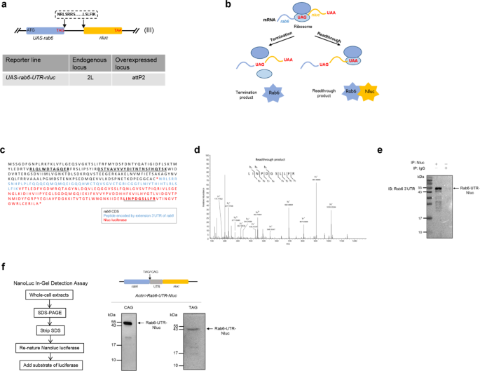
flies. RESULTS AN _IN VIVO_ GAIN-OF-FUNCTION REPORTER FLY LINE CAN SENSITIVELY DETECT TRANSLATIONAL READTHROUGH We wished to measure stop codon readthrough in flies across developmental
stages of their life cycle. We initially chose to measure readthrough using a candidate gene, _rab6_, which had been identified as undergoing moderately elevated readthrough rates in a
ribosome profiling study of fly embryos12. We decided to use a gain-of-function reporter15,16, since this approach would be able to detect low levels of readthrough. The gene for Nanoluc
luciferase – Nluc –17, missing its start codon, was cloned immediately downstream of _rab6_ complete with its native stop codon and 3′ UTR, but missing the second, in-frame, stop codon (Fig.
1a). In our reporter, translation past the native UAG stop codon would result in functional Nanoluc luciferase enzyme, which could be detected using commercially available reagents (Fig.
1b). We were able to identify by tandem mass spectrometry a peptide derived from Nluc in flies expressing the reporter (Fig. 1c,d). To further confirm that readthrough was occurring, we
raised a polyclonal antibody to a peptide coded by the 3′ UTR of _rab6_, and verified that a protein band of the appropriate size was identified by Western blot (Fig. 1e). To verify Nluc
protein was actually the product of translation past the stop codon, not alternative initiation of _nluc_ translation bypassing the stop codon, we performed an “in-gel” Nanoluc luciferase
assay capable of detecting functional enzyme on an SDS-PAGE separated whole-cell lysate (see Methods). We could detect functional Nluc as the major band corresponding to the size of the
Rab6-translated-3′ UTR-Nluc gene product, comparable to a control Rab6-Nluc fusion protein where the native TAG stop codon of _rab6_ had been replaced by CAG, corresponding to a glutamine
residue (Fig. 1f). Although some smaller bands were visible, they comprised a minority of the total signal, and may have represented alternate initiation or breakdown products. Taken
together, these data confirm that Nluc was expressed in reporter flies as a result of stop codon readthrough. THE STOP CODON OF REPORTER FLIES IS RECOGNIZED AS A PREMATURE TERMINATION CODON
We wished to use our reporter system to measure relative readthrough rates in a semi-quantitative way. To control for differential expression of the reporter, both the
UAS-_rab6-UTR-_STOP-_nluc_ reporter and UAS-_gfp_ were driven by Gal4 (see Methods and Fig. S1a). Comparison of the GFP and Nluc ratios would allow for relative quantitation of stop codon
readthrough18,19. To investigate potential cellular mediators of readthrough in our reporter system, we crossed our reporter fly line with a focused sub-library of UAS-RNAi fly lines from
the Harvard _Drosophila_ RNAi Screening Centre (DRSC) Resource20 at Tsinghua. Knock-down was driven via an actin-promoter except where knockdown of the target gene via an actin-driver
promoter was lethal, in which case knock-down was driven by _BM-40-SPARC-Gal4_ and _Cg-Gal4_. We chose to define “hits” if Nluc/GFP signal was either two-fold decreased or increased compared
with control. We screened a total of 615 candidate genes, and identified 90 potential regulators of _rab6_ readthrough (Fig. S1b). To eliminate _rab6_-specific hits, we rescreened the hits
with a second reporter line encoding a second readthrough candidate _rps20_12. Testing knock-down of these hits with the rps20 readthrough reporter identified 25 genes, which showed similar
readthrough phenotypes when knocked down in both _rab6_ and _rps20_ lines (Fig. S1c–f and Dataset S1). Only one candidate, the nonsense-mediated mRNA decay (NMD) gene _upf1_21,22, showed
increased Nluc/GFP in _rab6_ and _rps20_ reporters when knocked down (Fig. S2a). Stop codons can be classified as ‘native’ or ‘premature’, and mechanisms of both termination and readthrough
vary and depend on how the stop codon is recognized by the cellular machinery22. Nonsense-mediated mRNA decay, is an important surveillance mechanism for monitoring of transcripts encoding
PTCs, and prevents their translation by rapidly degrading such transcripts. Our identification of _upf1_ as a potential mediator of readthrough in our screen suggested that our reporter
constructs were recognized as coding premature termination codons (PTCs). However, systematic investigation of Upf substrates in yeast and mammalian cells suggest that apparently normal
mRNAs without the classical features of PTC-containing transcripts may also be targeted for degradation23,24,25. Was the native _rab6_ transcript a non-canonical Upf substrate? Knock-down of
_upf1_ led to increased abundance of reporter mRNA (Fig. 2a), but the stability of native _rab6_ mRNA was not significantly affected by _upf1_ knockdown (Fig. 2b). These data suggested that
rather than representing native gene readthrough, readthrough rates measured in our reporter flies represented variation in PTC suppression. To further confirm whether our reporters
measured PTC readthrough, we knocked down two other NMD-associated factors, _smg5_ and _upf_326. Knockdown of both factors increased reporter abundance (Fig. 2c,d), as well as Nluc/GFP
measurements (Fig. S2b), verifying that the reporter constructs, but not the native genes are subject to NMD, and that relative readthrough rates measured using the reporters would represent
PTC readthrough. PTC READTHROUGH IS REGULATED IN A DEVELOPMENTAL STAGE-DEPENDENT MANNER Since the initially-designed reporters were expressed as two constructs, and only one, expressing
Nluc, but not the other expressing GFP was subject to NMD, it was possible that our apparent readthrough rates may be skewed, and that some of the hits from the RNAi screen represented
interference with NMD and not readthrough _per se_. We therefore constructed a single-fusion reporter construct, representing _egfp-rab6-TAG-UTR-nluc_ (Fig. 3a). Activity from the reporter
was detected only when both Nluc and Gal4 were expressed (Fig. 3b), verifying its specificity and absence of background signal. The Nluc signal from whole-cell extract was derived almost
entirely from a protein representing the correct molecular weight for the fusion product, which was strongly supportive that the reporter represented readthrough (Fig. 3c). Furthermore,
immunoprecipitating and blotting against GFP identified two proteins with sizes representing normally the terminated translation, as well as a minority product (<1%) representing the
extended polypeptide that would result from stop codon readthrough (Fig. 3d). To determine whether the new reporter was also subject to NMD, and therefore whether readthrough represented PTC
suppression, we raised flies on cycloheximide or DMSO27. Cycloheximide treatment can stabilize mRNA transcripts subject to NMD28,29. Consistent with our other reporters, the new fusion
reporter, but not native _rab6_ also appeared to be subject to NMD (Fig. 3e,f). Having constructed a reporter that would measure relative PTC readthrough rates, we wanted to determine
whether levels of readthrough in our PTC reporter varied by development stage (Fig. S3). We measured Nluc/GFP through larval development (Fig. 3g,h) and also when adults were hatched at day
0, through day 50 of adult life (Fig. 3i). In immature flies, readthrough increased, peaking at the pupa stage (Fig. 3h). In adult flies, readthrough increased from the newly hatched stage
through to day 20, but decreased thereafter, although staying higher than the newly hatched adult (Fig. 3i), suggesting that increase in readthrough was not due to aging _per se_. These
results were confirmed with a second fusion _egfp-rps20-TAA-UTR-nluc_ reporter (Fig. S4). CERTAIN TYPES OF NEURONS UNDERGO HIGHER RATES OF STOP CODON READTHROUGH To test the spatial
variation of PTC-mediated readthrough, we constructed a series of reporter flies expressing the fusion reporters, driven by tissue-specific promoters. There were clear differences in levels
of readthrough by larval tissue, with the highest PTC readthrough in brain tissue (Fig. 4a). To further confirm whether neuronal or glial cells were responsible for the high readthrough
rates, we compared stop codon (PTC) readthrough in neurons with those in glia, by expression of the reporters in those tissues specifically. Neurons exhibited higher readthrough than glial
cells (Figs. 4b, S5). Could differences in NMD and hence reporter mRNA stability explain this observation? In both neurons and glia, the reporter was subject was to NMD, but to a similar
extent (Fig. 4c,d), and _nluc_ transcript abundance was similar in both tissues (Fig. 4e), suggesting that neither tissue variation in NMD or transcript abundance were sufficient to explain
the observed differences between glia and neurons. However, measured Nluc activity, corrected by GFP (Fig. 4b) or _nluc_ transcript abundance (Fig. 4f) was higher in neurons than glia.
Furthermore, we measured Nluc abundance, as well as the size of the Nluc-containing polypeptide by the Nluc in-gel assay, in reporters expressed in neurons or glia. For similar loading of
either total protein or non-extended reporter (as measured by blotting against Actin or GFP respectively), there was substantially more Nluc, at a size corresponding to the readthrough
product, in neurons compared with glia (Fig. 4g). These data confirm that PTC-readthrough in fly neurons occurred at higher rates than in fly glia. Finally, we wished to investigate whether
PTC readthrough rates varied by type of neuron. Using neurotransmitter-specific expression of the _egfp-rab6-TAG-UTR-nluc_ reporter, we observed that readthrough rates were highest in
cholinergic neurons, at more than twice the relative rate seen across neurons as a whole. By contrast, relative readthrough rates in dopaminergic and serotoninergic neurons was significantly
lower than the pan-neuronal rates (Fig. 4h), suggesting that even within fly brains, PTC-readthrough rates varied substantially. DISCUSSION HIGHER READTHROUGH RATES IN OLD FLIES IS NOT
ASSOCIATED WITH AGING Most studies on errors in gene translation have been on single-celled organisms, such as bacteria and yeast18,30,31,32,33 and therefore the relationship between
translational error and development or anatomy has received limited attention. Mistranslation increases in response to stress, for example viral infection or oxidative damage34, and
therefore it has been proposed that increased mistranslation over time may result in “error catastrophe” and may be one of the causes of aging35,36,37. Experimental evidence for this has
been limited. Measuring fidelity of translation of polyU transcripts _in vitro_ in brain homogenates from young and old rats, Filion and Laughrea failed to identify increased error rates
with old age38. A more recent study found a strong positive correlation between fidelity of translation of the first and second codon and longevity in rodents39, suggesting that although
mistranslation may not increase in old age, getting to old age may require high fidelity translation. However, of note, in that study, there was no correlation between stop codon readthrough
and longevity39. In our study, whilst we found that older adult flies had higher rates of readthrough than newly hatched adults (Fig. 3i), this increase in readthrough did not increase
further in very old flies (Fig. 3i), suggesting that some other regulatory factor than ‘old age’ may be responsible for the observation. SPECIFIC NEURONAL TYPES ARE RESPONSIBLE FOR HIGHER
RATE OF PTC READTHROUGH IN CNS Studies of tissue-specific translational regulation are still in their infancy40. A recent study measured the translatome by ribosome profiling of fly muscle
through tissue-specific expression of tagged ribosomes41. Mutations in Rpl38 resulted in profound patterning defects due to tissue-specific expression of Rpl38 and its role in translation of
Homeobox mRNAs42. CNS-specific expression of a mutated tRNA resulted in neurodegeneration in mice43, again verifying that components and regulation of the translation apparatus can be
expressed in a tissue-specific manner. A recent study observed high rates of native stop codon readthrough in mouse brains, although differences in readthrough rates between neurons and glia
were not apparent10. A recent seminal study examined olfaction in _Drosophila_44. In _D. sechellia_, a key olfactory receptor gene, _Ir75a_ has a premature termination codon, and the gene
had been classified as a “pseudogene”. However, Prieto-Godino and colleagues elegantly demonstrated substantial translation of full-length, functional Ir75a protein, due to PTC-readthrough.
Intriguingly, this phenomenon was also observed in _D. melanogaster_, in which _Ir75a_ has no PTC, suggesting a conserved, generalizable mechanism of PTC readthrough in olfactory neurons in
flies44. Similar to us, they observed that there was no substantial readthrough in fly glia, suggesting that our findings are not restricted to our candidate reporters. Our study extends
these findings by measurement of relative PTC-readthrough in several fly tissues, and confirmed that highest readthrough rates were observed in neurons, but more specifically, in a subset of
neurons, being highest in cholinergic, followed by glutaminergic neurons, and relatively low rates of readthrough in dopaminergic neurons. The difference in PTC-readthrough between fly glia
and neurons was not due to differences in NMD activity. Further mechanistic studies are needed to explain the high readthrough in specific _Drosophila_ neurons, perhaps by differential
expression studies in different neuronal subtypes. READTHROUGH MAY REPRESENT AN ALTERNATIVE MECHANISM OF PTC TRANSCRIPT RESCUE Readthrough of the stop codon in our reporters mechanistically
represented readthrough of a PTC (Fig. 2) and as such we cannot determine whether canonical readthrough rates also vary by developmental stage and anatomical site. In mammals, a stop codon
is usually labelled as premature if it is located more than 50 nucleotides upstream of the last exon-exon junction21,45. However, in _Drosophila_, as well as yeast, the definition of a PTC
occurs independently of exon-exon junctions46, and may include other elements for classification, such as “faux” 3′ UTRs47. Long 3′ UTRs are permissive for targeting of non-PTC-containing
transcripts as substrates of Upf1 and NMD48,49,50. It is therefore likely that by incorporating the _nluc_ gene in the extended 3′ UTRs of our reporter constructs, despite conserving all
other elements of the stop codon context, the ‘native’ stop codons of _rab6_ and _rps20_ were re-classified as PTCs by the _Drosophila_ cellular machinery. Despite their potentially
disastrous consequences and association with pathology51, premature termination codon-containing transcripts are surprisingly common. Alternative splicing of mRNA can result in a high
frequency of PTC-containing transcripts52. And one study estimated that the typical human individual codes for approximately 100 nonsense- (PTC-containing) gene variants, of which 20 would
be homozygous53. Since translation of proteins abnormally truncated due to the presence of a premature termination codon could result in protein misfolding, all eukaryotic cells have evolved
nonsense-mediated mRNA degradation as a quality control mechanism to target PTC-containing transcripts for destruction22,45. However, NMD is not 100% efficient, a significant proportion of
PTC-containing transcripts escape NMD54. Remarkably, a recent comparison of homozygous PTC-containing genes in humans with their homozygous “wild-type” counterparts showed that mRNA and
protein abundance were similar in the two groups, suggesting that escape of PTC-containing transcripts from NMD-mediated degradation may be the norm rather than the exception55. The
Upf-mediated NMD pathway has regulatory functions beyond surveillance of PTC-containing mRNA and may itself be repressed in a developmental manner22,56. Bioinformatic predictions have
suggested that neural tissue may be particularly susceptible to mistranslation-misfolded protein-induced damage57,58. It was therefore surprising that a brain-specific microRNA, miR-128,
actively repressed NMD59, and that this downregulation of NMD represented a switch that triggered neuronal development60. Potential mechanisms for rescue pathways for nonsense-containing
transcripts that do not rely on NMD, include RNA editing61, alternative splicing, truncated proteins bypassing the stop codon, and readthrough of the stop codon55. Our data supports a model
in which neuronal cells may still be protected from the potentially deleterious effects of translating PTC-containing transcripts, despite downregulation of NMD62 via enhanced readthrough of
PTC-containing mRNAs. MATERIALS AND METHODS FLY GENETICS Standard fly husbandry techniques and genetic methodologies were used to assess transgenes in the progeny of crosses, construct
intermediate fly lines and obtain flies of the required genotypes for each experiment63. The Gal4-UAS binary expression system was used to drive transgene expression with temporal and
spatial control64. Flies were cultured at 25 °C, and anesthetized by CO2 prior to use in experiments. Fly strains used in this study are listed in Table S1. Intermediate strains were
constructed using these strains. GENERATION OF NLUC-TAGGED READTHROUGH REPORTER TRANSGENIC LINES To obtain _UAS-RpS20-TAA-Nluc_, _UAS-Rab6-TAG-Nluc_ and _UAS-Rab6-CAG-Nluc_ lines, the
relevant _rps20-TAA-nluc_, _rab6-TAG-nluc_ and _rab6-CAG-nluc_ were separately cloned into vector pVALIUM10-roe using Gateway recombination. RNA extracted from S2 cells (a kind gift from Dr.
Gong Cheng) was used to synthesize cDNA (Bio-Rad, cat#1708890). Coding sequences of _rps20_, _rab6_ and their following extension _3_′ _UTR_12 were PCR-amplified from cDNA template with
primers adding _attB_ site at the 5′ termini of the ORF. In order to increase the expression of the transgenes in _Drosophila_, a 5′ UTR element _Syn21_65 and the Kozak sequence CAAAATG (the
start codon underlined)66 were added to the coding sequence. _nluc_ was codon optimized by GENEWIZ (Nanjing), and both start codon and stop codon were removed, and this _nluc_ gene was
inserted before the second in-frame stop codon of the extension 3′ UTR to acquire a fusion protein. Simultaneously, _attB_ site was added to the 3′ termini of the modified fusion protein for
subsquent Gateway cloning. Additionally, _rab6-CAG-nluc_ was constructed by site directed mutagenesis using _rab6-TAG-nluc_ as template. To construct _UAS-eGFP-Rab6-TAG-Nluc_ and
_UAS-eGFP-RpS20-TAA-Nluc_, _egfp_, missing its stop codon, was PCR-amplified and fused with _rab6-TAG-nluc_ and _rps20-TAA-nluc_, respectively by overlap PCR. The PCR products were purified
by gel extraction (cwbiotech, cat#CW2302M) and recombined into vector pDONR221 (Life Technologies, cat#12536017) using Gateway BP Clonase (Life Technologies, cat#11789020). Then the entry
clones were recombined with destination vector pVALIUM10-roe using Gateway LR clonase (Life Technologies, cat#12538120). The final plasmids UAS-RpS20-TAA-Nluc, UAS-Rab6-TAG-Nluc,
UAS-eGFP-Rab6-TAG-Nluc, UAS-eGFP-RpS20-TAA-Nluc and UAS-Rab6-CAG were sent to Tsinghua Fly Center to obtain transgenic fly lines20 by site-directed insertion. To overexpress protein on the
second chromosome, entry clone was integrated into attP40 loci, while the overexpressed protein on the third chromosome was obtained by integrating into attP2 loci67. All the primers used in
transgenic fly lines construction are listed in Table S2. LUCIFERASE-GFP ASSAYS Luciferase was measured using the NanoGlo Luciferase Assay Kit (Promega, cat#N1120). Euthanized flies were
collected in 200 μl of NB buffer (150 mM NaCl, 50 mM Tris-HCl pH 7.5, 2 mM EDTA, 0.1% NP-40) with addition of protease inhibitor cocktail (Biotool, cat#B14003), and homogenized with a
96-well plate multiple homogenizer (Burkard Scientific, BAMH-96). Homogenized samples were centrifuged at 20,000 rcf (4 °C) to pellet the larval remains. For measuring readthrough level
(Nluc/GFP), 30 μl of each sample supernatant was transferred to a white-walled 96-well plate (Costar, cat#3922), an equal volume of Promega Luciferase Reagent was added to each well and
incubated in the dark for 5 min. Another 30 μl of each sample supernatant was correspondingly transferred to a black-walled 96-well plate (Corning, cat#3925), and an equal volume of NB
buffer was added to each well. Luminescence and fluorescence signal were measured by Fluoroskan Ascent FL (Thermo Scientific). IMMUNOPRECIPITATION OF NLUC For western blot (Fig. 1e), Nluc
luciferase was immunoprecipitated by addition of 12 μg of rabbit polyclonal anti-Nluc IgG (a generous gift from Lance Encell, Promega) or normal rabbit IgG as a negative control (Cell
Signaling Technology, cat#2729) respectively, to concentrated fly lysates, and incubated overnight at 4 °C to form the immune complex. The immune complex was captured by Protein A/G Plus
Agarose (Pierce, cat#26146) and eluted by heating the resin with SDS sample buffer. For mass spectrometry (Fig. 1d), readthrough product of _rab6_ was enriched by a Pierce Direct IP Kit
(Pierce, cat#26148). For each enrichment, 10 μg of rabbit polyclonal anti-Nluc IgG (Promega) was coupled to AminoLink Plus Coupling Resin, and concentrated cell lysate was incubated with the
resin overnight at 4 °C to form the immune complex. The enriched readthrough product was eluted by a neutral pH elution buffer (Thermo Scientific, cat#21027). IMMUNOPRECIPITATION OF
EGFP-TAGGED READTHROUGH REPORTER For western blot (Fig. 3d), eGFP-tagged readthrough reporter was immunoprecipitated by addition of 10 μg of mouse monoclonal anti-GFP IgG (Roche,
cat#11814460001) or normal mouse IgG as a negative control (Proteintech, cat#66360-3-Ig) respectively, to concentrated fly lysates, and incubated overnight at 4 °C to form the immune
complex. The immune complex was captured by Protein A/G Plus Agarose (Pierce, cat#26146) and eluted by heating the resin with SDS sample buffer. NANOLUC IN-GEL DETECTION ASSAY The protocol
from the Promega technical manual (https://www.promega.com/-/media/files/resources/protocols/technical-manuals/500/nano-glo-in-gel-detection-system-technical-manual.pdf?la=en) was followed.
Briefly, cell lysates were resolved on 15% SDS-PAGE. The gel was extracted from its casing and transferred to an appropriate tray. SDS was stripped from the gel by washing three times with
25% isopropanol in water (20 min each). NanoLuc was re-natured with three water washes (20 min each). The gel was developed with Furimazime (Promega, cat#N1120)/TBST (50 mM Tris-HCl pH 7.6,
150 mM NaCl, 0.05% Tween 20, 25 uM Furimazine). The gel image was captured on a white reflective background by Chemidoc XRS + (Bio-Rad). WESTERN BLOT Western bolt was performed using
standard methods. Rabbit polyclonal antibody to _Drosophila_ Rab6 3′ UTR was acquired by immunizing the New Zealand rabbit with peptide NRLSRRSNHPLPLFC by GenScript company (Nanjing).
Readthrough product of _rab6_ (Fig. 1e) was detected using rabbit anti-Rab6 3′ UTR antibody (2 μg/ml, GenScript) and revealed with Clean-Blot IP Detection Reagent (Thermo Scientific,
cat#21230) and ECL Western Blotting Substrate (Pierce, cat#32106). For eGFP-tagged reporter Western blots (Fig. 3d), elution was detected using 1:2500 dilution of monoclonal rat anti-GFP
antibody (chromotek, RRID: AB_10773374), followed by a 1:5000 dilution of goat anti-rat IgG-HRP secondary antibody (easybio, cat#BE0109). Expression of reporter from input was detected by
mouse anti-GFP IgG (Roche, cat#11814460001), followed by a 1:5000 dilution of goat anti-mouse IgG-HRP secondary antibody (cwbiotech, cat#CW0102). Loading control of neural cells (Fig. 4g)
was using 1:2000 dilution of anti-insect beta Actin mouse antibody (cmctag, cat#AT0008), followed by a 1:5000 dilution of goat anti-mouse IgG-HRP secondary antibody, or 1:2500 dilution of
monoclonal rat anti-GFP antibody as mentioned before. MASS SPECTROMETRY Readthrough product of _rab6_ was enriched by a developed IP method. After electrophoresis, SDS PAGE was stained by
Imperial Protein Stain (Thermo Scientific, cat#24615), and the gel between 43 kDa to 55 kDa were cut, digested with trypsin, analyzed by Tsinghua Protein Chemistry Facility. CANDIDATE
FORWARD GENETIC SCREEN Readthrough reporter fly line _Actin_ > _Rab6-TAG-Nluc_ or _Actin_ > _RpS20-TAA-Nluc_ was crossed with a set of _UAS-RNAi_ fly lines (Tsinghua Fly Center) to
knockdown target genes. Reporter fly line was crossed with _w_11,18, and readthrough level (Nluc/GFP) of progeny was as control. _BM-_4_0-SPARC_ > _RpS20-TAA-Nluc_ and _Cg_ >
_Rab6-TAG-Nluc_ reporter fly lines were utilized when knockdown target genes by _actin_ > _Gal4_ resulted in growth deficiency. For each knockdown genotype, 8–12 wandering L3 larvae were
collected to measure readthrough level. Readthrough level of control was normalized to 1, and for each knockdown genotype, relative readthrough fold to control was calculated. DEVELOPMENTAL
STAGE ASSAY Parent transgenic reporter line _y v sc; UAS-RpS20-TAA-Nluc_ (II) or _y v sc; UAS-Rab6-TAG-Nluc_ (III) and an actin driven Gal4 line _w; Sp/CyO; act-FO-Gal4 UAS-GFP/TM6B_ was
crossed to express the readthrough reporter ubiquitously in the whole body of progeny. To eliminate the effect by variation in NMD efficiency, reporter line _y v sc; UAS-eGFP-RpS20-TAA-Nluc_
(II) or _y v sc; UAS-eGFP-Rab6-TAG-Nluc_ (III) was crossed with _y w; Adv/CyO; actin-Gal4/TM6B_. After 6 hours of crossing, parent flies were removed to ensure the majority of the progenies
were in the same developmental stage. Cell lysates were flash frozen by liquid nitrogen and stored at −80 °C until all the samples of different developmental stages had been collected.
Readthrough value was measured in whole cell lysate as above. For embryo collection, standard apple juice agar plates were supplemented with fresh baker yeast paste, and collection cages
were placed on the plates. Parent flies lay eggs on the plates for 4 hours. 0–4 hr embryos were collected from the agar plate using a small paintbrush. For aged flies collection, newly
hatched male and female flies were sorted into independent groups and cultured them in standard environmental conditions with a 12:12 hr light dark cycle. During the experimental period,
flies were transferred to new vials containing fresh food every 2–3 days68. After 40 days of adult life, flies started to die and living flies would be transferred to fresh food every day to
avoid the flies sticking to the food in the old vial. PROTEIN EXTRACTION FROM _DROSOPHILA_ EMBRYOS Collected embryos were washed gently in a collection basket (Corning, cat#352350). The
base of the basket was dried by a paper tissue, and the basket was transferred to a container with 50% commercial bleach solution. Incubated for 5 min with gentle, periodic stirring to
remove the chorionic membrane of the embryos. The dechorionated embryos became hydrophobic and floated on the surface of the bleach solution. Transferred the basket to a new container with
deionized water, washed for 2 min and repeated twice. After wash, about 30 embryos of each independent sample were transferred to a 1.5 ml eppendorf tube containing 200 ul of ice-cold NB
lysis buffer (protease inhibitor cocktail was added) and homogenized by a cordless motor (Kimble, cat#749540-0000). Homogenized samples were centrifuged at 20,000 rcf (4 °C) to pellet the
larval remains, and supernatant avoiding the upper lipid layer was transferred to a new 1.5 ml eppendorf tube. PROTEIN EXTRACTION FROM DIFFERENT _DROSOPHILA_ ORGANS OR TISSUES Expression of
readthrough reporter in wing disc was driven by _rn-Gal4_, gut by _crq-Gal4_, salivary gland by _He-Gal4_, fat body by _Cg-Gl4_, neuron by _elav-Gal4_, glial cells by _repo-Gal4_ and
_Nrv2-Gal4_. Different larval organs or tissues were dissected and collected in 100 μl of NB lysis buffer with addition of protease inhibitor cocktail, homogenized by a cordless motor.
Homogenized samples were centrifuged at 20,000 rcf (4 °C) to pellet the larval remains, and supernatant avoiding the upper lipid layer was transferred to a new 1.5 ml eppendorf tube. The
final cleared cell lysates were flash frozen by liquid nitrogen and stored at −80 °C until all the samples of different _Drosophila_ tissues or organs had been collected. ANTIBIOTICS
TREATMENT L3 larvae were transferred to standard fly food containing 500 μg/ml cycloheximide (MedChemExpress, cat#HY-12320)27 or DMSO for 24 h. Afterwards, total RNA were extracted for
real-time RT-PCR. REAL-TIME QUANTITATIVE RT-PCR Approximate 10–20 wandering L3 larvae were collected and RNA was extracted by TransZol Up (TransGen Biotech, cat#ET111-01). DNase I (TransGen
Biotech, cat#GD201-01) was used to digest genomic DNA. cDNA was synthesized with iScript Reverse Transcription kit (Bio-Rad, cat#1708890) and iTaq Universal SYBR Green Supermix (Bio-Rad,
cat#1725120) was used for quantitative PCR. Analysis was performed in a CFX96 Real-Time PCR Detection System (Bio-Rad). _rp49_ was used as a reference gene69,70. All PCR reactions were
performed in biological triplicate. Primers used were: _rp49_-For: 5′-GGCCCAAGATCGTGAAGAAG-3′; _rp49_-Rev: 5′-ATTTGTGCGACAGCTTAGCATATC-3′; _upf1_-For: 5′-ACTTCCGGTTCGCACATCAT-3′; _upf1_-Rev:
5′-CTTCCACTGTTCCTGGTCCC-3′; _upf3_-For: 5′- ATGCTCCCTTCCAGTGCTTC-3′; _upf3_-Rev: 5′- CCGCTTGATGAACTCCTGGT-3′; _smg5_-For: 5′- GCTTTTTGACTGGCTGCGAA-3′; _smg5_-Rev: 5′-
ACCAGAGAATCACGCACGTT-3′; _nluc_-For: 5′-GATCATCCCCTACGAGGGCT-3′; _nluc_-Rev: 5′-GTCGATCATGTTGGGGGTCA-3′; _rab6_-3′UTR-For: 5′-ATCCAACCATCCTCTCCCCC-3′; _rab6_-3′UTR-Rev:
5′-GCAGATCCGGCCAGTACATA-3′; _rps20_-3′UTR-For: 5′- ATCATCGACTTGCACTCGCC-3′; _rps20_-3′UTR-Rev: 5′- GCACGCCAAACTTTTCGAGG-3′. QUANTITATION AND STATISTICAL ANALYSIS Most of experiments were
performed at least three times on separate days (that is, independent experiments). Statistical analysis and graphic representation were performed with Graphpad Prism software. Two-tailed
non-parametric Mann-Whitney tests were used in Fig. S2A (data did not pass normality tests), otherwise means between two sample sets were compared by unpaired, two-directional Student’s
_t_-tests in the rest of comparisons. *_P_ < 0.05, **_P_ < 0.01, ***_P_ < 0.001, ****_P_ < 0.0001 were considered statistically significant results. REFERENCES * Dever, T. E.
& Green, R. The elongation, termination, and recycling phases of translation in eukaryotes. _Cold Spring Harb Perspect Biol_ 4, a013706, https://doi.org/10.1101/cshperspect.a013706
(2012). Article CAS PubMed PubMed Central Google Scholar * Brown, A., Shao, S., Murray, J., Hegde, R. S. & Ramakrishnan, V. Structural basis for stop codon recognition in
eukaryotes. _Nature_ 524, 493–496, https://doi.org/10.1038/nature14896 (2015). Article ADS CAS PubMed PubMed Central Google Scholar * von der Haar, T. & Tuite, M. F. Regulated
translational bypass of stop codons in yeast. _Trends Microbiol_ 15, 78–86, https://doi.org/10.1016/j.tim.2006.12.002 (2007). Article CAS PubMed Google Scholar * Firth, A. E. &
Brierley, I. Non-canonical translation in RNA viruses. _J Gen Virol_ 93, 1385–1409, https://doi.org/10.1099/vir.0.042499-0 (2012). Article CAS PubMed PubMed Central Google Scholar *
Halfmann, R. _et al_. Prions are a common mechanism for phenotypic inheritance in wild yeasts. _Nature_ 482, 363–368, https://doi.org/10.1038/nature10875 (2012). Article ADS CAS PubMed
PubMed Central Google Scholar * True, H. L. & Lindquist, S. L. A yeast prion provides a mechanism for genetic variation and phenotypic diversity. _Nature_ 407, 477–483 (2000). Article
ADS CAS Google Scholar * Steneberg, P. & Samakovlis, C. A novel stop codon readthrough mechanism produces functional Headcase protein in Drosophila trachea. _EMBO Rep_ 2, 593–597,
https://doi.org/10.1093/embo-reports/kve128 (2001). Article CAS PubMed PubMed Central Google Scholar * Eswarappa, S. M. _et al_. Programmed translational readthrough generates
antiangiogenic VEGF-Ax. _Cell_ 157, 1605–1618, https://doi.org/10.1016/j.cell.2014.04.033 (2014). Article CAS PubMed PubMed Central Google Scholar * Loughran, G. _et al_. Evidence of
efficient stop codon readthrough in four mammalian genes. _Nucleic Acids Res_ 42, 8928–8938, https://doi.org/10.1093/nar/gku608 (2014). Article CAS PubMed PubMed Central Google Scholar
* Sapkota, D. _et al_. Cell-Type-Specific Profiling of Alternative Translation Identifies Regulated Protein Isoform Variation in the Mouse Brain. _Cell reports_ 26, 594–607 e597,
https://doi.org/10.1016/j.celrep.2018.12.077 (2019). Article CAS PubMed PubMed Central Google Scholar * Jungreis, I. _et al_. Evidence of abundant stop codon readthrough in Drosophila
and other metazoa. _Genome Res_ 21, 2096–2113, https://doi.org/10.1101/gr.119974.110 (2011). Article CAS PubMed PubMed Central Google Scholar * Dunn, J. G., Foo, C. K., Belletier, N.
G., Gavis, E. R. & Weissman, J. S. Ribosome profiling reveals pervasive and regulated stop codon readthrough in Drosophila melanogaster. _Elife_ 2, e01179,
https://doi.org/10.7554/eLife.01179 (2013). Article PubMed PubMed Central Google Scholar * Li, C. & Zhang, J. Stop-codon read-through arises largely from molecular errors and is
generally nonadaptive. _PLoS Genet_ 15, e1008141, https://doi.org/10.1371/journal.pgen.1008141 (2019). Article CAS PubMed PubMed Central Google Scholar * Arribere, J. A. _et al_.
Translation readthrough mitigation. _Nature_ 534, 719–723, https://doi.org/10.1038/nature18308 (2016). Article ADS CAS PubMed PubMed Central Google Scholar * Su, H. W. _et al_. The
essential mycobacterial amidotransferase GatCAB is a modulator of specific translational fidelity. _Nat Microbiol_ 1, 16147, https://doi.org/10.1038/nmicrobiol.2016.147 (2016). Article CAS
PubMed Google Scholar * Ribas de Pouplana, L., Santos, M. A., Zhu, J. H., Farabaugh, P. J. & Javid, B. Protein mistranslation: friend or foe? _Trends in biochemical sciences_ 39,
355–362, https://doi.org/10.1016/j.tibs.2014.06.002 (2014). Article CAS PubMed Google Scholar * Hall, M. P. _et al_. Engineered luciferase reporter from a deep sea shrimp utilizing a
novel imidazopyrazinone substrate. _ACS chemical biology_ 7, 1848–1857, https://doi.org/10.1021/cb3002478 (2012). Article CAS PubMed PubMed Central Google Scholar * Javid, B. _et al_.
Mycobacterial mistranslation is necessary and sufficient for rifampicin phenotypic resistance. _Proc Natl Acad Sci USA_ 111, 1132–1137, https://doi.org/10.1073/pnas.1317580111 (2014).
Article ADS CAS PubMed Google Scholar * Kramer, E. B. & Farabaugh, P. J. The frequency of translational misreading errors in E. coli is largely determined by tRNA competition. _RNA_
13, 87–96, rna.294907 (2007). * Ni, J. Q. _et al_. Vector and parameters for targeted transgenic RNA interference in Drosophila melanogaster. _Nat Methods_ 5, 49–51,
https://doi.org/10.1038/nmeth1146 (2008). Article CAS PubMed Google Scholar * Gupta, P. & Li, Y. R. Upf proteins: highly conserved factors involved in nonsense mRNA mediated decay.
_Mol Biol Rep_ 45, 39–55, https://doi.org/10.1007/s11033-017-4139-7 (2018). Article CAS PubMed Google Scholar * He, F. & Jacobson, A. Nonsense-Mediated mRNA Decay: Degradation of
Defective Transcripts Is Only Part of the Story. _Annual review of genetics_ 49, 339–366, https://doi.org/10.1146/annurev-genet-112414-054639 (2015). Article CAS PubMed PubMed Central
Google Scholar * Hurt, J. A., Robertson, A. D. & Burge, C. B. Global analyses of UPF1 binding and function reveal expanded scope of nonsense-mediated mRNA decay. _Genome Res_ 23,
1636–1650, https://doi.org/10.1101/gr.157354.113 (2013). Article CAS PubMed PubMed Central Google Scholar * Tani, H. _et al_. Identification of hundreds of novel UPF1 target transcripts
by direct determination of whole transcriptome stability. _RNA Biol_ 9, 1370–1379, https://doi.org/10.4161/rna.22360 (2012). Article CAS PubMed PubMed Central Google Scholar * Guan, Q.
_et al_. Impact of nonsense-mediated mRNA decay on the global expression profile of budding yeast. _PLoS Genet_ 2, e203, https://doi.org/10.1371/journal.pgen.0020203 (2006). Article CAS
PubMed PubMed Central Google Scholar * Avery, P. _et al_. Drosophila Upf1 and Upf2 loss of function inhibits cell growth and causes animal death in a Upf3-independent manner. _RNA_ 17,
624–638, https://doi.org/10.1261/rna.2404211 (2011). Article CAS PubMed PubMed Central Google Scholar * King-Jones, K., Charles, J. P., Lam, G. & Thummel, C. S. The ecdysone-induced
DHR4 orphan nuclear receptor coordinates growth and maturation in Drosophila. _Cell_ 121, 773–784, https://doi.org/10.1016/j.cell.2005.03.030 (2005). Article CAS PubMed Google Scholar *
Colak, D., Ji, S. J., Porse, B. T. & Jaffrey, S. R. Regulation of axon guidance by compartmentalized nonsense-mediated mRNA decay. _Cell_ 153, 1252–1265,
https://doi.org/10.1016/j.cell.2013.04.056 (2013). Article CAS PubMed PubMed Central Google Scholar * Inoue, K. _et al_. Molecular mechanism for distinct neurological phenotypes
conveyed by allelic truncating mutations. _Nat Genet_ 36, 361–369, https://doi.org/10.1038/ng1322 (2004). Article CAS PubMed Google Scholar * Meyerovich, M., Mamou, G. & Ben-Yehuda,
S. Visualizing high error levels during gene expression in living bacterial cells. _Proc Natl Acad Sci USA_ 107, 11543-11548, doi:0912989107 (2010). * Miranda, I. _et al_. Candida albicans
CUG mistranslation is a mechanism to create cell surface variation. _MBio_ 4, https://doi.org/10.1128/mBio.00285-13 (2013). * Ruan, B. _et al_. Quality control despite mistranslation caused
by an ambiguous genetic code. _Proc Natl Acad Sci USA_ 105, 16502–16507 (2008). 0809179105. Article ADS CAS Google Scholar * Schwartz, M. H. & Pan, T. Temperature dependent
mistranslation in a hyperthermophile adapts proteins to lower temperatures. _Nucleic Acids Res_ 44, 294–303, https://doi.org/10.1093/nar/gkv1379 (2016). Article CAS PubMed Google Scholar
* Netzer, N. _et al_. Innate immune and chemically triggered oxidative stress modifies translational fidelity. _Nature_ 462, 522–526, nature08576 (2009). * Orgel, L. E. The maintenance of
the accuracy of protein synthesis and its relevance to ageing. _Proceedings of the National Academy of Sciences_ 49, 517–521 (1963). Article ADS CAS Google Scholar * Orgel, L. E. The
maintenance of the accuracy of protein synthesis and its relevance to ageing: a correction. _Proceedings of the National Academy of Sciences_ 67, 1476–1476 (1970). Article ADS CAS Google
Scholar * Orgel, L. E. Ageing of clones of mammalian cells. _Nature_ 243, 441 (1973). Article ADS CAS Google Scholar * Filion, A. M. & Laughrea, M. Translation fidelity in the aging
mammal: studies with an accurate _in vitro_ system on aged rats. _Mechanisms of ageing and development_ 29, 125–142 (1985). Article CAS Google Scholar * Ke, Z. _et al_. Translation
fidelity coevolves with longevity. _Aging cell_ 16, 988–993 (2017). Article CAS Google Scholar * Shi, Z. & Barna, M. Translating the genome in time and space: specialized ribosomes,
RNA regulons, and RNA-binding proteins. _Annu Rev Cell Dev Biol_ 31, 31–54, https://doi.org/10.1146/annurev-cellbio-100814-125346 (2015). Article CAS PubMed Google Scholar * Chen, X.
& Dickman, D. Development of a tissue-specific ribosome profiling approach in Drosophila enables genome-wide evaluation of translational adaptations. _PLoS Genet_ 13, e1007117,
https://doi.org/10.1371/journal.pgen.1007117 (2017). Article CAS PubMed PubMed Central Google Scholar * Kondrashov, N. _et al_. Ribosome-Mediated Specificity in Hox mRNA Translation and
Vertebrate Tissue Patterning. _Cell_ 145, 383–397, https://doi.org/10.1016/j.cell.2011.03.028 (2011). Article CAS PubMed PubMed Central Google Scholar * Ishimura, R. _et al_. RNA
function. Ribosome stalling induced by mutation of a CNS-specific tRNA causes neurodegeneration. _Science_ 345, 455–459, https://doi.org/10.1126/science.1249749 (2014). Article ADS CAS
PubMed PubMed Central Google Scholar * Prieto-Godino, L. L. _et al_. Olfactory receptor pseudo-pseudogenes. _Nature_ 539, 93–97, https://doi.org/10.1038/nature19824 (2016). Article ADS
CAS PubMed PubMed Central Google Scholar * Nicholson, P. & Muhlemann, O. Cutting the nonsense: the degradation of PTC-containing mRNAs. _Biochem Soc Trans_ 38, 1615–1620,
https://doi.org/10.1042/BST0381615 (2010). Article CAS PubMed Google Scholar * Gatfield, D., Unterholzner, L., Ciccarelli, F. D., Bork, P. & Izaurralde, E. Nonsense-mediated mRNA
decay in Drosophila: at the intersection of the yeast and mammalian pathways. _EMBO J_ 22, 3960–3970, https://doi.org/10.1093/emboj/cdg371 (2003). Article CAS PubMed PubMed Central
Google Scholar * Amrani, N. _et al_. A faux 3′-UTR promotes aberrant termination and triggers nonsense-mediated mRNA decay. _Nature_ 432, 112–118, https://doi.org/10.1038/nature03060
(2004). Article ADS CAS PubMed Google Scholar * Hogg, J. R. & Goff, S. P. Upf1 senses 3′ UTR length to potentiate mRNA decay. _Cell_ 143, 379–389,
https://doi.org/10.1016/j.cell.2010.10.005 (2010). Article CAS PubMed PubMed Central Google Scholar * Kurosaki, T. & Maquat, L. E. Rules that govern UPF1 binding to mRNA 3′ UTRs.
_Proc Natl Acad Sci USA_ 110, 3357–3362, https://doi.org/10.1073/pnas.1219908110 (2013). Article ADS PubMed Google Scholar * Kebaara, B. W. & Atkin, A. L. Long 3′-UTRs target
wild-type mRNAs for nonsense-mediated mRNA decay in Saccharomyces cerevisiae. _Nucleic Acids Res_ 37, 2771–2778, https://doi.org/10.1093/nar/gkp146 (2009). Article CAS PubMed PubMed
Central Google Scholar * Mort, M., Ivanov, D., Cooper, D. N. & Chuzhanova, N. A. A meta-analysis of nonsense mutations causing human genetic disease. _Hum Mutat_ 29, 1037–1047,
https://doi.org/10.1002/humu.20763 (2008). Article CAS PubMed Google Scholar * McGlincy, N. J. & Smith, C. W. Alternative splicing resulting in nonsense-mediated mRNA decay: what is
the meaning of nonsense? _Trends in biochemical sciences_ 33, 385–393, https://doi.org/10.1016/j.tibs.2008.06.001 (2008). Article CAS PubMed Google Scholar * MacArthur, D. G. _et al_. A
systematic survey of loss-of-function variants in human protein-coding genes. _Science_ 335, 823–828, https://doi.org/10.1126/science.1215040 (2012). Article ADS CAS PubMed PubMed
Central Google Scholar * Isken, O. & Maquat, L. E. Quality control of eukaryotic mRNA: safeguarding cells from abnormal mRNA function. _Genes Dev_ 21, 1833–1856,
https://doi.org/10.1101/gad.1566807 (2007). Article CAS PubMed Google Scholar * Jagannathan, S. & Bradley, R. K. Translational plasticity facilitates the accumulation of nonsense
genetic variants in the human population. _Genome Res_ 26, 1639–1650, https://doi.org/10.1101/gr.205070.116 (2016). Article CAS PubMed PubMed Central Google Scholar * Feng, Q.,
Jagannathan, S. & Bradley, R. K. The RNA Surveillance Factor UPF1 Represses Myogenesis via Its E3 Ubiquitin Ligase Activity. _Mol Cell_ 67, 239–251 e236,
https://doi.org/10.1016/j.molcel.2017.05.034 (2017). Article CAS PubMed PubMed Central Google Scholar * Drummond, D. A. & Wilke, C. O. Mistranslation-induced protein misfolding as a
dominant constraint on coding-sequence evolution. _Cell_ 134, 341–352, https://doi.org/10.1016/j.cell.2008.05.042 S0092-8674(08)00705-8 [pii] (2008). * Drummond, D. A. & Wilke, C. O.
The evolutionary consequences of erroneous protein synthesis. _Nat Rev Genet_ 10, 715–724, nrg2662 (2009). * Bruno, I. G. _et al_. Identification of a microRNA that activates gene expression
by repressing nonsense-mediated RNA decay. _Mol Cell_ 42, 500–510, https://doi.org/10.1016/j.molcel.2011.04.018 (2011). Article CAS PubMed PubMed Central Google Scholar * Lou, C. H.
_et al_. Posttranscriptional control of the stem cell and neurogenic programs by the nonsense-mediated RNA decay pathway. _Cell reports_ 6, 748–764,
https://doi.org/10.1016/j.celrep.2014.01.028 (2014). Article CAS PubMed PubMed Central Google Scholar * Duan, Y., Dou, S., Luo, S., Zhang, H. & Lu, J. Adaptation of A-to-I RNA
editing in Drosophila. _PLoS Genet_ 13, e1006648, https://doi.org/10.1371/journal.pgen.1006648 (2017). Article CAS PubMed PubMed Central Google Scholar * Jaffrey, S. R. & Wilkinson,
M. F. Nonsense-mediated RNA decay in the brain: emerging modulator of neural development and disease. _Nat Rev Neurosci_ 19, 715–728, https://doi.org/10.1038/s41583-018-0079-z (2018).
Article CAS PubMed PubMed Central Google Scholar * Roote, J. & Prokop, A. How to design a genetic mating scheme: a basic training package for Drosophila genetics. _G3 (Bethesda)_ 3,
353–358, https://doi.org/10.1534/g3.112.004820 (2013). Article Google Scholar * Brand, A. H. & Perrimon, N. Targeted gene expression as a means of altering cell fates and generating
dominant phenotypes. _development_ 118, 401–415 (1993). CAS PubMed Google Scholar * Pfeiffer, B. D., Truman, J. W. & Rubin, G. M. Using translational enhancers to increase transgene
expression in Drosophila. _Proc Natl Acad Sci USA_ 109, 6626–6631, https://doi.org/10.1073/pnas.1204520109 (2012). Article ADS PubMed Google Scholar * Cavener, D. R. Comparison of the
consensus sequence flanking translational start sites in Drosophila and vertebrates. _Nucleic acids research_ 15, 1353–1361 (1987). Article CAS Google Scholar * Markstein, M., Pitsouli,
C., Villalta, C., Celniker, S. E. & Perrimon, N. Exploiting position effects and the gypsy retrovirus insulator to engineer precisely expressed transgenes. _Nat Genet_ 40, 476–483,
https://doi.org/10.1038/ng.101 (2008). Article CAS PubMed PubMed Central Google Scholar * Linford, N. J., Bilgir, C., Ro, J. & Pletcher, S. D. Measurement of lifespan in Drosophila
melanogaster. _J Vis Exp_, https://doi.org/10.3791/50068 (2013). * Zang, Y. _et al_. Plasma membrane overgrowth causes fibrotic collagen accumulation and immune activation in Drosophila
adipocytes. _Elife_ 4, e07187, https://doi.org/10.7554/eLife.07187 (2015). Article CAS PubMed PubMed Central Google Scholar * Ponton, F., Chapuis, M. P., Pernice, M., Sword, G. A. &
Simpson, S. J. Evaluation of potential reference genes for reverse transcription-qPCR studies of physiological responses in Drosophila melanogaster. _J Insect Physiol_ 57, 840–850,
https://doi.org/10.1016/j.jinsphys.2011.03.014 (2011). Article CAS PubMed Google Scholar Download references ACKNOWLEDGEMENTS We thank Dr.Yi Zhong (_elav-Gal4 and repo-Gal4_) and Dr. Wei
Zhang (_Cha-Gal4_, _VGlut-Gal4_, _TH-Gal4_, _GAD1-Gal4_ and _Trh-Gal4_) for kindly sharing fly strains; Lance Encell, Paul Otto and Thomas Machleidt from Promega for generously providing
the anti-Nluc antibody and the protocol for the NanoLuc luciferase In-Gel Detection Assay; and the Tsinghua Fly Center for constructing transgenic _in vivo_ readthrough reporter fly lines.
This work was supported in part by start-up funds from Tsinghua University to BJ; and a grant from the National Science Foundation of China [31771600] to JCPP. BJ is an Investigator of the
Wellcome Trust [207487/B/17/Z]. The funders had no role in study design, data collection and analysis, decision to publish, or preparation of the manuscript. AUTHOR INFORMATION AUTHORS AND
AFFILIATIONS * Center for Global Health and Infectious Disease Research, Collaborative Innovation Center for the Diagnosis and Treatment of Infectious Diseases, Tsinghua University School of
Medicine, Beijing, 100084, China Yanan Chen, Zhuo Bi & Babak Javid * School of Life Sciences, Tsinghua University, Beijing, 100084, China Yanan Chen, Tianhui Sun, Zhuo Bi & Jose C.
Pastor-Pareja * Tsinghua University School of Medicine, Beijing, 100084, China Jian-Quan Ni * Tsinghua-Peking Joint Center for Life Sciences, Tsinghua University, Beijing, 100084, China Jose
C. Pastor-Pareja * Beijing Advanced Innovation Center in Structural Biology, Beijing, China Babak Javid Authors * Yanan Chen View author publications You can also search for this author
inPubMed Google Scholar * Tianhui Sun View author publications You can also search for this author inPubMed Google Scholar * Zhuo Bi View author publications You can also search for this
author inPubMed Google Scholar * Jian-Quan Ni View author publications You can also search for this author inPubMed Google Scholar * Jose C. Pastor-Pareja View author publications You can
also search for this author inPubMed Google Scholar * Babak Javid View author publications You can also search for this author inPubMed Google Scholar CONTRIBUTIONS B.J. conceived the
project. Y.N.C. and B.J. designed research. Y.N.C. performed the vast majority of experiments with assistance from T.H.S. and Z.B. J.Q.N. provided the platform for generation of fly lines.
J.C.P.P. and B.J. supervised research. Y.N.C. and B.J. wrote the manuscript with input from J.C.P.P. and other authors. CORRESPONDING AUTHORS Correspondence to Jose C. Pastor-Pareja or Babak
Javid. ETHICS DECLARATIONS COMPETING INTERESTS The authors declare no competing interests. ADDITIONAL INFORMATION PUBLISHER’S NOTE Springer Nature remains neutral with regard to
jurisdictional claims in published maps and institutional affiliations. SUPPLEMENTARY INFORMATION SUPPLEMENTARYFILE 2. SUPPLEMENTARY FIGURES RIGHTS AND PERMISSIONS OPEN ACCESS This article
is licensed under a Creative Commons Attribution 4.0 International License, which permits use, sharing, adaptation, distribution and reproduction in any medium or format, as long as you give
appropriate credit to the original author(s) and the source, provide a link to the Creative Commons license, and indicate if changes were made. The images or other third party material in
this article are included in the article’s Creative Commons license, unless indicated otherwise in a credit line to the material. If material is not included in the article’s Creative
Commons license and your intended use is not permitted by statutory regulation or exceeds the permitted use, you will need to obtain permission directly from the copyright holder. To view a
copy of this license, visit http://creativecommons.org/licenses/by/4.0/. Reprints and permissions ABOUT THIS ARTICLE CITE THIS ARTICLE Chen, Y., Sun, T., Bi, Z. _et al._ Premature
termination codon readthrough in _Drosophila_ varies in a developmental and tissue-specific manner. _Sci Rep_ 10, 8485 (2020). https://doi.org/10.1038/s41598-020-65348-8 Download citation *
Received: 03 January 2020 * Accepted: 31 March 2020 * Published: 22 May 2020 * DOI: https://doi.org/10.1038/s41598-020-65348-8 SHARE THIS ARTICLE Anyone you share the following link with
will be able to read this content: Get shareable link Sorry, a shareable link is not currently available for this article. Copy to clipboard Provided by the Springer Nature SharedIt
content-sharing initiative